
Nadia Iftikhar ( Department of Dermatology, Pakistan. )
Amer Ejaz ( Military Hospital, Rawalpindi. Department of Dermatology, Combined Military Hospital, Kharian Cantonment, Pakistan. )
Umar Aftab Butt ( Department of Dermatology, Pakistan. )
Salman Ali ( Department of Paediatrics, Pakistan. )
November 2009, Volume 59, Issue 11
Case Reports
Abstract
Aplasia cutis congenita is a rare skin condition characterized by the absence of localized or widespread areas of skin at birth. We are reporting a variant aplasia cutis congenita, which involved over 90% of the body surface area, which occurred in a baby born to a mother with pemphigus vulgaris who was on oral prednisolone and azathioprine. A case of extensive aplasia cutis congenita was seen and oral intake of azathioprine by the mother during pregnancy was suspected as an etiologic factor. The parents of the patient did not consent for biopsy or autopsy so the histopathological picture was not available and hence involvement of other systems could not be ascertained. Due, to financial constraints some of the investigations which might have helped in assigning the patient to a particular category of aplasia cutis such as karyotyping and CT scan brain could not be carried out.
Introduction
Aplasia cutis congenita (ACC) is a condition characterized by the absence of skin at birth. The defect can involve a small portion of the scalp or widespread areas. The most commonly involved site is the scalp. There is no racial or sex predilection. It is classified into nine groups based on the etiology and the clinical findings. Our patient suffered from isolated extensive ACC and was exposed in utero to both, azathioprine, and prednisolone. Azathioprine has been held responsible for an increase in congenital malformations although not specifically ACC and prednisolone is regarded safe in pregnancy.
Case Report
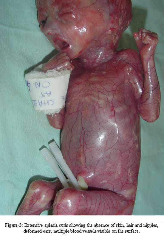
A new born baby boy was seen in the neonatal Intensive Care Unit (ICU) just after birth, who had almost complete absence of the skin except for some area around the nose (Figure-1) and on the buttocks (Figure-2) where skin had formed. The entire body was covered in a membrane which had numerous vessels visible on its surface. He had no nails or hair and nipples were not formed (Figure-3). He had micrognathia, his ears were malformed and his right testis was undescended. He was born with a very low birth weight 1.02 kg, fronto-occipital circumference was 26.5 cm (normal range 33- 38 cm) and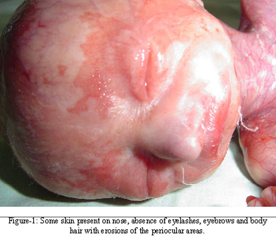
crown heel length was 37 cm (normal range 48- 53cm). He was nursed in an incubator but died on the 4th day of birth of neonatal sepsis in spite of supportive management, including antibiotic therapy.
He was born to non consanguineous parents by lower segment caesarian section at 38 weeks of gestation because the baby showed signs of intrauterine growth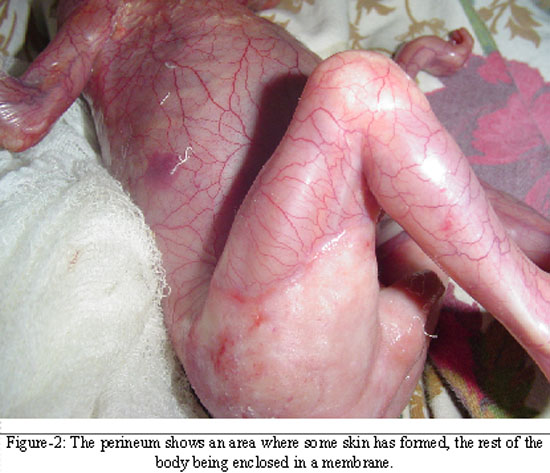
retardation. The mother had a history of two previous c-sections. Four other children were alive and healthy. There was no history of chicken pox or herpes zoster during pregnancy. The mother who was 26 years of age had a history of pemphigus vulgaris for the past 1 year, confirmed on histopathology and direct immunoflourescence. Her disease had been particularly difficult to control and she had been on tablet prednisolone 60 mg daily for the first 2 months of pregnancy. Gradual reduction was attempted but she developed fresh blisters when the dose was reduced below 30 mg daily, so 30mg/day had to be continued for the rest of her pregnancy. In addition, to prednisolone she was on azathioprine 150 mg daily (3mg/kg body weight) 6 weeks prior to and for the first 20 weeks of pregnancy. After delivery of the baby the dose of prednisolone was reduced gradually and her disease was controlled on 15 mg of prednisolone on alternate days.
Discussion
Pemphigus Vulgaris during pregnancy is very rare.1 Our case was further complicated by the birth of a neonate with extensive skin aplasia. Aplasia cutis congenita is a fairly common birth defect. What makes the case interesting is that this neonate was exposed to two drugs in utero, prednisolone before and throughout pregnancy and azathioprine 6 weeks before and for the first 20 weeks of pregnancy.
Corticosteroids, prednisone and prednisolone cross the placental barrier, but are metabolized by a placental 11- hydroxygenase and thus the foetus is exposed to only 10% of the maternal dose. Corticosteroids in pregnancy have caused cleft palate and lip in experimental animal models and low birth weight in humans.2 However, there is no evidence that prednisolone is teratogenic in humans.3 (FDA category B).
Azathioprine is an inhibitor of purine metabolism that decreases natural killer cell activity, antibody production, antibody dependant cellular cytotoxicity and cellular immune response such as interleukin 2 secretion.3 It is one of the most commonly used immunomodulatory drugs in the treatment of organ transplant rejection and in the treatment of various autoimmune disorders.
After absorption azathioprine is converted into its active metabolite 6 mercaptopurine (6-MP) and other metabolites. Azathioprine and mercaptopurine both cross the placenta and mercaptopurine can be measured in foetal blood. In theory the foetus is protected from the adverse effects of azathioprine in early pregnancy as its liver lacks the enzyme inosine pyrophosphorylase, that converts azathioprine into its active metabolites and only trace amounts of its metabolites are detected in foetal serum.4 Azathioprine is a FDA Category D drug, there is evidence of risk to human foetuses however it may be used in pregnancy if benefits outweigh risks.
Azathioprine teratogenicity has been demonstrated in animals (malformations include cleft palate, hydrocephalus, skeletal defects, ocular abnormalities and limb osteogenesis).4
The use of azathioprine in pregnancy is related to intrauterine growth retardation, an increased risk of stillbirth, preterm birth, and low birth weight, together with a 2.8% prevalence of malformations in the exposed.4-6 Another, study suggests that the risk of congenital malformations is dose dependant and varies from 3-9% in antenatally exposed infants.7
The malformations reported with azathioprine include microcephaly, myelomeningocele, thymic atrophy, adrenal hypoplasia and hypospadias.7 Haematologic abnormalities have also been reported.2 A child with aphakia and another with hypoplasia and dysplasia of the lungs and malformations of the urinary bladder and urethra has been reported.4 An infant born with preaxial polydactyly to a mother taking azathioprine throughout pregnancy is described.8
Francella et al6 evaluated the potential toxicity of 6-MP and found no statistically significant difference in spontaneous abortion rate, abortion as a result of a birth defect, major congenital malformations, neoplasia, or increased infections among male or female patients taking 6-MP compared with control subjects (relative risk, 0.85 [95% CI, 0.47-1.55]; P < 0.59). The authors concluded, on the basis of their findings, that 6-MP use before or at the time of conception or during pregnancy appear to be safe.
Extensive data, published on azathioprine treated recipients suggests that while there is a pattern of prematurity among the newborn, there has not been an increase in the incidence or pattern of specific malformations noted among the newborn.3 Other studies, suggest that standard doses of azathioprine does not increase the risk of congenital anomalies and azathioprine can be given safely throughout pregnancy.6
ACC has been reported with maternal use of methimazole, carbimazole, benzodiazepine, misoprostol, and valproate.7 To date, an association with azathioprine has not been reported.
Aplasia Cutis has also been described as a feature of a number of malformation syndromes (Type 9) including trisomy 13, the 4p-syndrome, Oculocerebrocutaneous syndrome, Johanson Blizzard syndrome and Focal facial dermal dysplasia but all of these syndromes are characterized by limited areas of aplasia cutis and have in addition other associated features which were absent in our patient.9 The Patau syndrome (trisomy 13) in addition to limited areas of aplasia cutis also has iris colobomas, cleft lip/ or palate, polydactyly, narrow and curved nails.9 The 4p- syndrome is associated with a broad nose, preauricular tags or pits.9 Affected individuals experience fits and are mentally retarded. The oculocerebro cutaneous syndrome is associated with eyelid colbomas, orbital cysts, skull defects, cerebral malformations and porencephaly.9 Johanson- Blizzard syndrome is associated with rectourogenital anomalies, thyroid dysfunction and microcephaly.9 Focal facial dermal dysplasia is associated with focal lesions on the face.9 Park et al described a male infant with extensive areas of aplasia cutis congenita in association with choanal atresia, syndactyly, imperforate anus and pulmonary hypoplasia.10 They could not identify any drug or malformation syndrome and the karyotype of the infant was normal. Unfortunately our patient could not be investigated extensively due to financial constraints.
Our patient had extensive ACC and it appeared to be an isolated finding. So we conclude that our patient is most likely type 8 ACC — as there was no evidence of limb reduction defects, epidermolysis bullosa, intrauterine infection or twin pregnancy, on the other hand, exposure to azathioprine at the time of conception and for the following 20 weeks could be responsible for this occurrence. We believe it is important to document this extensive skin aplasia and although a chance occurrence cannot be ruled out, the extensive and severe nature of the defect makes us wary and wonder if azathioprine really is an innocent bystander?
Conclusion
Our case is more likely associated with Type 8 ACC, which is the type associated with teratogens. Azathioprine could probably be the cause in this case, or it could be a chance occurrence. Type 1 although not associated with multiple abnormalities and has only localized lesions on the scalp; but as the investigations were not done, the abnormalities cannot be ruled out.
References
1.Goldberg NS, DeFeo G, Kirshenbaum N. Pemphigus Vulgaris and pregnancy: risk factors and recommendations. J Am Acad Dermatol 1993; 28: 877-9.
2.Janssen NM, Genta MC. The effects of immunosuppressive drugs and anti-inflammatory medications on fertility, pregnancy, and lactation. Arch Intern Med 2000; 160: 610-9.
3.Prevot A, Martini S, Guignard JP. In utero exposure to immunosuppressive drugs. Biol Neonate 2002; 81: 73-81.
4.Norgard B, Pedersen L, Fonager K, Rasmussen SN, Sorensen HT. Azathioprine, mercaptopurine and birth outcome: a population based cohort study. Alimentary Pharmacol Ther 2003; 17: 827-34.
5.Tendron A, Gouyon JB, Decramer S. In utero exposure to immunosuppressive drugs:experimental and clinical studies. Pediatr Nephrol 2002; 17:121-30.
6.Francella A, Dyan A, Bodian C, Rubin P, Chapman M, Present DH. The safety of 6-mercaptopurine for childbearing patients with inflammatory bowel disease: a retrospective cohort study. Gastroenterology 2003; 124: 9-17.
7.Zukiene J, Zalgeviciene V, Rizgeliene R. The influence of azathioprine on the osteogenesis of the limbs. Medicina (Kaunas) 2003; 39: 584-8.
8.Miniero R, Tardivo I, Curtoni ES, Segoloni GP, La Rocca E, Nino A, et al. Pregnancy after renal transplantation in Italian patients: focus on fetal outcome. J Nephrol 2002; 15: 626-32.
9.Sparker MK, Garcia-Gonzalez E, Sanchez LT. Sclerosing and Atrophying conditions. In Lawrence A. Schachner, Ronald C. Hansen, editors. Pediatric Dermatology 3rd edition. p. 793-5.
10.Park MS, Hahn SH, Hong CH, Kim JS, Kim HS. Extensive form of aplasia cutis congenita: a new syndrome? Journal of Med Genet 1998; 35: 609-11.
Journal of the Pakistan Medical Association has agreed to receive and publish manuscripts in accordance with the principles of the following committees: