
Mohammad Tahir ( Department of Anatomy, University of Health Sciences, Lahore. )
Khalid Perwez Lone ( Department of Physiology, University of Health Sciences, Lahore. )
Nadia Ahmad ( M. Phil Research Scholar, University of Health Sciences, Lahore. )
July 2016, Volume 66, Issue 7
Original Article
Abstract
Objective: To observe the ameliorating effect by methanolic extract of pomegranate peel in acetaminophen-induced hepatotoxicity.
Methods: The randomised controlled study was conducted from July 2013 to June 2014 at the University of Health Sciences, Lahore, Pakistan, and comprised rats that were randomly divided into three equal groups. Control group A was given normal saline (5ml/kg), whereas group B and C were given 750mg/kg acetaminophen intraperitoneally dissolved in normal saline (5ml/kg) on 1st day of experiment. From Day 2 till day 14, group A and B were given distilled water (5ml/kg), while group C was given 50mg/kg methanolic extract of pomegranate peel dissolved in distilled water (5ml/kg) orally. On day 15, blood was collected through cardiac puncture, and livers were removed and processed for histological examination.
Results: There were 24 rats weighing 175±25gm each. Each group had 8(33.3%) rats. Mean liver aspartate aminotransferase at the end of the experiment in groups A, B and C were 97.88±19.45, 148.25±16.48 and 96.13±17.95U/L, while alanine transaminase levels were 51.50±15.38, 96.75±10.91 and 49.63±12.08 U/L (p<0.05 each) On histological examination of group B, the normal hepatic architecture was distorted with loss of classically arranged hepatic cords. Vascular congestion was present with centrilobular necrosis, marked by pyknotic nuclei and vacuoles.
Conclusion: Acetaminophen is hepatotoxic and methanolic extract of pomegranate peel ameliorated the hepatic picture probably because of its antioxidant properties.
Keywords: Acetaminophen, Pomegranate peel, Hepatotoxicity. (JPMA 66: 859; 2016)
Introduction
Acetaminophen, commonly known as paracetamol, is one of the most widely used over-the-counter analgesic and antipyretic for both children and adults.1 Acetaminophen, an aniline, non-steroidal anti-inflammatory drug (NSAID) belongs to the para-amino phenol class and is an active metabolite of two other anilines, phenacetin and acetanilide which were both found to metabolise to paracetamol and P-phenatidine or aniline, respectively.2
Acetaminophen, when taken in toxic doses, can cause hepatic necrosis, nephrotoxicity and death in humans and experimental animal alike.3 Adult conventional dose of acetaminophen is 0.5-1gm every four to six hours with a maximum daily dose of 4gm.4 In children the dose depends on age and body weight which is less than 40mg/kg in 24 hours.5
More than 90% of acetaminophen, taken in therapeutic doses, is metabolised in the liver to phenolic glucuronide and sulfate by glucuronyltransferases and sulfotransferases and is subsequently excreted in the urine.6 About 2% of the residual acetaminophen is excreted unchanged in the urine and about 5% to 10% gets metabolised by the cytochrome P450 to N-acetyl-p-benzoquinoneimine (NAPQI).6
With acetaminophen overdose, glucuronyltransferases and sulfotransferases get saturated, diverting the drug to be metabolised by cytochrome P450. This generates NAPQI in amounts that can deplete glutathione, which, if not replenished, results in the accumulation of NAPQI in the hepatocytes.7 Secondary to NAPQI-induced glutathione depletion and oxidative stress, lipid peroxidation takes place which leads to irreversible cell membrane injury and, hence, cell death.7,8
Pomegranate (PunicaGranatum L.) plant extract from its different parts possess antioxidant activity, which is highest among many foods.9 Chemically, the plant includes diverse polyphenol antioxidants, primarily ellagic acid and punicalagin.10 Peels of pomegranate contain high content of polyphenols such as condensed tannins and proanthocyanidins, anthocyanins (delphinidin, cyanidin and pelargonidin 3-glucosides and 3, 5-diglucosides) and flavinoids which are referred to as antioxidants.11 These compounds are known for their properties in scavenging free radicals and inhibiting lipid peroxidation.11 Dried fruit peel is reported to exert diverse pharmacological functions with antioxidant activity and is used for diarrhoea and for the treatment of respiratory and urinary tract infections.12 Parts or extracts of different parts of this plant had been used as anti-cancer, anti-bacterial, anti-diarrhoeal, anti-fungal and anti-ulcer remedy.13 Peel also has antifertility,14 hepatoprotective,15 cytotoxic,16 and hypoglycaemic activities.17 Studies report that the peel in particular possesses relatively higher antioxidant activity than other parts of pomegranate and, therefore, is a rich source of natural antioxidants.11 Since hepatoprotective effect of methanolic extract of pomegranate peel (MEPP) had never been tried on acetaminophen-induced hepatotoxicity, the present study was designed to observe the acetaminophen-induced histological and functional changes in the liver of rat and the effect of MEPP on these changes.
Materials and Methods
The randomised controlled study was conducted from July 2013 to June 2014 at the University of Health Sciences, Lahore, Pakistan, and comprised healthy male albino Wistar rats.
The animals were divided through random balloting into three equal groups A, B, and C and were individually housed in stainless steel cages with wood shavings on the floor. The animals were fed on standard rat diet and water ad libitum. The animals were kept at 23±2°C and humidity (50±5%). The photoperiod was controlled at 12 hours. The experiment was started 4 days after acclimatisation of the animals.
Group A, control, was given 5ml/kg normal saline intraperitoneally (IP) on the day 1 and then 5ml/kg distilled water orally from day 2 till day 14; groups B and C were given 750 mg/kg acetaminophen IP dissolved in 5ml/kg normal saline on day 1 of experiment. From day 2 till day 14, group B was given 5ml/kg distilled water, while group C was also given 50 mg/kg MEPP dissolved in 5ml/kg distilled water orally. On day 15, blood was drawn through cardiac puncture and serum was kept at -80ºC for the assessment of liver enzymes aspartate aminotransferase (AST) and alanine transaminase (ALT). The animals were then sacrificed under chloroform anaesthesia and the livers were removed, weighed and small pieces of 3-5mm were excised and prepared for histological examination. Data were statistically analysed using SPSS 20. Comparison of variables was made using analysis of variance (ANOVA) followed by Post Hoc Tukey test. P<0.05 was considered statistically significant.
Results
There were 24 rats weighing 175±25gm each. Each group had 8(33.3%) rats. Mean liver AST at the end of the experiment in groups A, B and C were 97.88±19.45, 148.25±16.48 and 96.13±17.95U/L, while ALT levels were 51.50±15.38, 96.75±10.91 and 49.63±12.08 U/L (Table)
Histologically, normal hepatolobular characteristics were seen in control animals (Figure-1)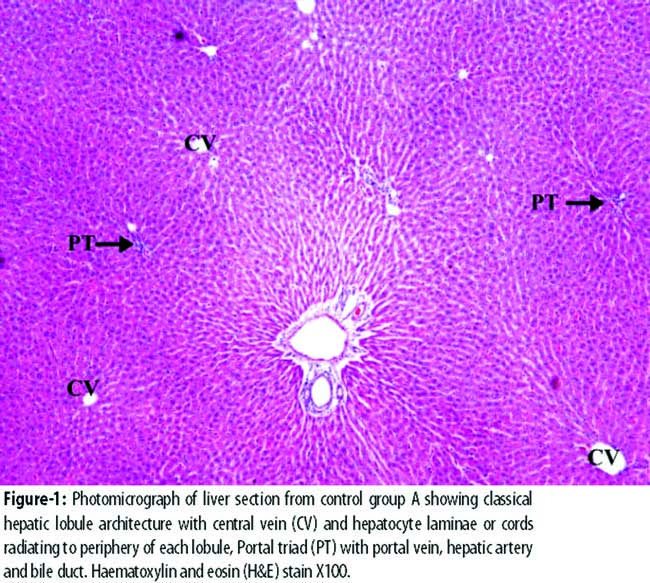
In the histological preparations from group B, general hepatic architecture was disrupted with loss of classical arrangement of hepatic cords. The cell boundary of hepatocytes was not clearly defined. Cytoplasm contained large number of vacuoles pushing the nucleus to one side. The nuclei were darkly stained, indicating pyknotic changes along with clumping of nuclear material. Nucleoli were not clearly seen in many hepatocytes. Necrosis was present in group B as evident by pyknosis. The liver parenchyma showed vascular congestion (Figure-2)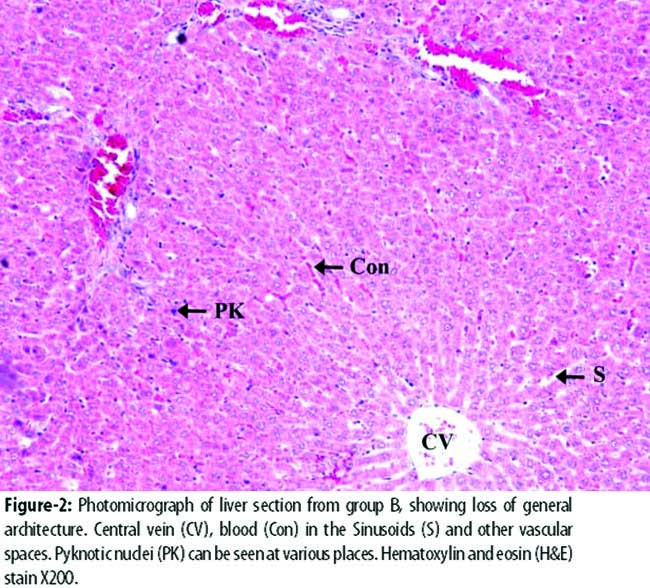
In group C, general architecture of liver parenchyma was restored. There was hardly any evidence of damage to the organ; no vascular congestion, necrosis or inflammation was observed. Signs of regeneration were present in group C as evident by cytoplasmic eosinophilia, prominent nucleoli, multinucleation and two cells thick liver cords (Figure-3)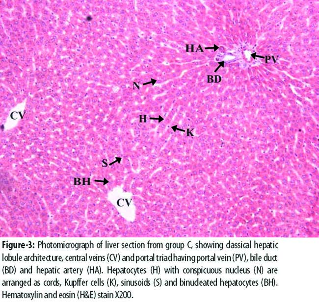
similar to those seen in control group A.
Discussion
The results showed that acetaminophen was toxic to liver as indicated by various histological changes and biochemical parameters. There are several reports showing that acetaminophen administration increased the lipid peroxidation, disturbing cell membrane integrity and releasing the liver enzymes into circulation.18-21 In the present investigation, it was observed that, in group B, serum levels of ALT and AST were significantly increased compared to those in group A (p<0.001); this was indicative of damage to the hepatocyte plasmalemma as reported earlier.18-21
Histological examination of the liver preparations in the current study showed that the normal architecture of liver in group B was deranged and hepatocytes showed swelling (Figure-2). This loss of classical arrangement of hepatocytes due to ballooning was also previously reported after acetaminophen treatment of albino rats.22,23 In the current experiment, MEPP restored the general architecture of liver in group C (Figure-3). The current results corroborate previous study16 in which administration of a single dose of 2g/kg of carbon tetrachloride (CCl4) caused disintegration and degradation of liver cell architecture and treatment with MEPP showed normal lobular pattern with well-formed polygonal hepatocytes having conspicuous nuclei. Vacuoles were observed within the hepatocytes in the present histological preparations of the liver from group B which was indicative of degenerating hepatocytes and/or an increased deposition of fat.24 It has been reported that the toxic metabolite of acetaminophen bound covalently to cell macromolecules; NAPQI, the toxic metabolite of acetaminophen was reported to be metabolised by cytochrome P-450 in liver and subsequently detoxified.24
Hepatotoxicity had been reported to result as a mark of oxidative stress and free radical production.24 It is also suggested that acetaminophen is responsible for hepatotoxicity in the current experiment by increasing oxidative stress in animals of group B and protection was provided by MEPP since the extract contained strong antioxidants and polyphenols such as condensed tannins and proanthocyanidins, anthocyanins (delphinidin, cyanidin and pelargonidin 3-glucosides and 3, 5-diglucosides) and flavinoids.11 It appears that the ingredients present in MEPP restored hepatotoxic changes by decreasing the oxidative stress-induced toxicity of acetaminophen (Figure-3).
Evidences of regeneration were present in group C which was treated with MEPP (Figure-3). Literature reports that increased cytoplasmic eosinophilia, multinucleated hepatocytes (mostly binucleated), prominent nucleoli and two cell thick liver cords seen in group C animals are suggestive of regeneration.25 Therefore, we can hypothesise that MEPP restored the toxic effects of acetaminophen when given orally. However, further detailed studies are needed to establish and confirm these effects.
Conclusion
The study showed acetaminophen-induced hepatotoxicity in rats as manifested by histological and functional changes; total loss of hepatic architecture with centrilobular hepatic necrosis, fatty changes, vacuolisation, loss of cell boundaries and elevation in levels of AST and ALT. However, treatment with MEPP reversed these changes, showing its ameliorating effects. MEPP deserves further intensive study as a protective and preventive agent since it has more potential being a rich source of antioxidants.
Acknowledgements
We are grateful to University of Health Sciences (UHS), Lahore, for financial support, and to the staff of Animal House and Anatomy Department for animal handling and experimentation.
Disclosure: There had been no disclosure.
Conflict of Interest: There is no conflict of interest.
Funding Source: University of Health Sciences, Lahore.
References
1. Laura PJ, Philip RM, Jack AH. Acetaminophen-Induced Hepatotoxicity. Drug Metab Dispos 2003; 31: 1499-506.
2. Toussaint K, Yang XC, Zielinski MA, Reigle KL, Sacavage SD, Nagar S, et al. What do we (not) know about how Paracetamol (acetaminophen) works? J Clin Pharm Ther 2010; 35: 617-38.
3. Ray SD, Mumaw VR, Raje RR, Faris MW. Protection of acetaminophen induced hepatocellular apoptosis and necrosis by cholesteryl hemisuccinol pretreatment. J Pharmacol Exp Ther 1996; 279: 1470-83.
4. Forte JS. Paracetamol: Safety versus Toxicity. The chronic Ill 2002; 1: 12-6.
5. Jefferies S, Saxena M, Young P. Paracetamol in critical illness: a review. Crit Care Resusc 2012; 14: 74-80.
6. Black M. Acetaminophen hepatotoxicity. Gastroenterology 1980; 78: 382-92.
7. Slitt AML, Dominick PK, Roberts JC, Cohen SD. Effect of ribose cysteine pretreatment on hepatic and renal acetaminophen metabolite formation and glutathinone depletion. Basic Clin Pharmacol Toxicol 2005; 96: 487-94.
8. Olaleye MT, Rocha BT. Acetaminophen-induced Liver damage in mice: Effects of some Medicinal plants on the oxidative defense system. Exp Toxicol Pathol 2008; 59: 319-27.
9. Guo C, Yang J, Wei J, Li Y, Xu J, Jiang Y. Antioxidant activities of peel, pulp and seed fractions of common fruits as determined by FRAP assay. Nutr Res 2003; 23: 1719-26.
10. Aviram M, Rosenblat M, Gaitini D, Nitecki S, Hoffman A, Dornfeld L, et al. Pomegranate juice consumption for 3 years by patients with carotid artery stenosis reduces common carotid intima-media thickness, blood pressure and LDL oxidation. Am J Clin Nutr 2004; 23: 423-33.
11. Wang Z, Pan Z, Ma H, Atungulu GG. Extract of Phenolics From Pomegranate Peels. Open Food Sci J 2011; 5: 17-25.
12. Ahmad MM, Ali SE. Protective effect of pomegranate peel ethanol extract against ferric nitrilotriacetate induced renal oxidative damage in rats. J Cell Mol Biol 2010; 7 & 8: 35-43.
13. Gurpreet K, Zoobi J, Mohammad A, Alam MS. Punicagranatum (pomegranate) flower extract possess potent antioxidant activity and abrogates Fe-NTA induced hepatotoxicity in mice. Food Chem Toxicol 2006; 44: 984-93.
14. Gujraj ML, Varma DR, Sareen KN. Oral contraceptives. Part 1. Preliminary observations on the antifertility effects of some indigenous drugs. Indian J Med Res 1960; 48: 46-51.
15. Murthy KN, Jayaprakasha GK, Singh RP. Studies on antioxidant activities of pomegranate peel extract using in vivo models. J Agri Food Chem 2002; 50: 4791-5.
16. KulkarniAP, Mahal HS, Kapoor S, Aradhya SM. In vitro studies on the binding, antioxidant, and cytotoxic actions of punicalagin. J Agric Food Chem 2007; 55: 1491-500.
17. Hontecillas R, O\\\'Shea M, Einerhand A, Diguardo M, Bassaganya-Riera J. Activation of PPAR gamma and alpha by punicic acid ameliorates glucose tolerance and suppresses obesity-related inflammation. J Am Coll Nutr 2009; 28: 184-95.
18. Olaleye MT, Rocha BT. Acetaminophen-induced liver damage in mice: effects of some medicinal plants on the oxidative defence system. Exp Toxicol Pathol 2008; 59:319-27.
19. Mladenovic D, Radosavljevic T, Ninkovic M, Vuc-evic D, Jesic-Vukic´evic R, Todorovic V. Liver antioxidant capacity in the early phase of acute paracetamol-induced liver injury in mice. Food Chem Toxicol 2009; 47: 866-70.
20. Sun J, Ando Y, Ahlbory-Dieker D, Schnackenberg LK, Yang X, Greenhaw J, et al. Systems Biology Investigation to Discover Metabolic Biomarkers of Acetaminophen-Induced Hepatic Injury Using Integrated Transcriptomics and Metabolomics. J Mol Biomark Diagn 2013; S1: 002.
21. Martin-Murphya BV, Holta MP, Jua C. The role of damage associated molecular pattern molecules in acetaminophen-induced liver injury in mice. Toxicol Lett 2010; 192: 387-94.
22. Abdel-Zehr AO, Abdel-Hady RH, Mahmoud MM, Farrag MM. The potential protective role of alpha-lipoic acid against acetaminophen-induced hepatic and renal damage. Toxicology 2008; 243: 261-70.
23. Gopalakrishnan S, Kalaiarasi T. Hepatoprotective activity of the ethanolic extract of the fruits of cucumistrigonusroxb. Int J Pharm Pharm Sci 2013; 5: 268-72.
24. Fromentry B. Bridging the gap between old and new concepts in drug-induced liver injury. Clin Res Hepatol Gastroenterol 2013; 31: 6-9.
25. Moneim AEA, Othman MS, Mohmoud SM, El-Dei KM. Pomegranate peel attenuates aluminum-induced hepatorenal toxicity. Toxicol Mech Meth 2013; 23: 624-33.
Journal of the Pakistan Medical Association has agreed to receive and publish manuscripts in accordance with the principles of the following committees: