

Aalia Nazir ( Department of Physics, The Islamia University of Bahawalpur, Pakistan. )
Saeed Ahmad Buzdar ( Department of Physics, The Islamia University of Bahawalpur, Pakistan. )
Muhammad Afzal Rao ( Department of Physics, The Islamia University of Bahawalpur, Pakistan. )
June 2010, Volume 60, Issue 6
Original Article

Abstract
Objective: This study focuses on the diverse effects of MRI parameters on image quality using widely available imaging pulse sequences.
Methods: A tissue equivalent gel system has been used for Magnetic Resonance Imaging by using Xylenol orange dye with Fricke-Benzoic solution which was formulated by Kelly R.G (1998). The gel system was radiated while using 6MV photons from Varian Clinic Linear Accelerator.
Results: The qualitative analysis include the effect of TR and TE on signal nonuniformity according to which the escalating repetition time gives a uniform signal having the average values calculated are 0.76%, 0.83%, 1.43% and 1.89% for the conventional spin echo (CSE), fast spin echo (FSE), gradient recalled echo (GRE) and fluid attenuated inversion recovery (FLAIR) respectively. FLAIR showed contradictory results due to longitudinal relaxation of the signal. This is because, at the longer value of inversion time (TI), the signal from simple fluids is nulled, thus reducing signal intensity.
Conclusion: The evaluation of signal uniformity for different pulse sequences demonstrates that the repetition time (TR) affects the signal homogeneity as it maintains image quality for CSE and FSE. However, a careful selection is required for FLAIR due to its sensitive behaviour for image uniformity (JPMA 60:470; 2010).
Introduction
For over 40 years ferrous sulphate (Fricke) solution had been used for radiation dosimetry.1,2 Gore et al,3 in 1984, proposed combining the system with magnetic resonance imaging to make possible three dimensional dosimetry. In gel Dosimetry, the phantom itself is the detector, thus providing the ability to measure the true three dimensional dose distributions. This is a unique property when compared to other dosimeters (TLDs, diodes, ionization chambers and film).4 In Fricke gels, Fe2+ ions in ferrous sulphate solutions are usually dispersed throughout a gel matrix. Fe2+ ions are converted to Fe3+ ions with a corresponding change in paramagnetic properties5 hence used to make an MRI image of the dose distribution. Several basic experimental studies were performed on Fricke gels investigating the effect of the gelling substances6,7 and with chelating agents such as Xylenol orange to reduce diffusion in the dosimeter gels.8,9 A relation was developed between dose, sensitivity and ferrous sulphate concentration10 and the relaxation mechanism at different field strengths.9,11 It was shown that by adding a metal ion indicator such as xylenol orange induces color changes12 in the gel upon irradiation, thus enable it to be scanned optically.13 They also alter the absorption spectra of the Fricke gels so that irradiated gels gives visible colour development.12,14
Material and Methods
FBX Gel preparation:
A number of different gel systems have been proposed for gel dosimetry, but agarose15-20 and gelatin (usually 300 BLOOM porcine) are the gels of choice. In our study, Ferrous Benzoic Xylenol Orange (FBX) gel was prepared using Gelatine (from bovine skin, Type B), Ferrous Ammonium sulphate (Aldrich Ammonium Iron (II) Sulphate Hexahydrate, 99% A.C.S Reagent), Sulphuric acid (Sigma Aldrich), Xylenol orange tetrasodium salt (Sigma Aldrich) and Benzoic Acid (Sigma Aldrich) formulated in 1998 by Kelly RG and others.21
The stock solution was prepared by mixing 5mM of Benzoic Acid, ImM of Xylenol Orange and 25mM of Sulphuric acid in a one litre volumetric flask, kept at room temperature. The gel preparation began with the addition of 40gm of gelatin in 700ml of distilled water of 25mM of Sulphuric acid which was preheated to ~40°C using a hot plate stirrer. The gelatin was dissolved in the gel by constant slow stirring for 30 minutes. In another beaker 0.1mM of ferrous sulphate was dissolved in 100ml of benzoic acid Xylenol orange stock solution and then added to liquid gelatin. Final volume of one litre of gel was completed by adding 25mM of sulphuric acid. The gel was prepared at room temperature allowed to be exposed to air throughout its preparation. This was because response of Fricke gel dosimeter is dependent upon the initial concentrations of oxygen present in the solution. The gel phantom was stored in a pyrex glass bottle kept at 5°C.
Gel Irradiation and MRI scanning:
The gel was irradiated using 6MV photons from a Varian Clinic 600C Linear accelerator, to a maximum dose of 20GY with a 5×5cm2 field size at 95.5cm SSD (Source to surface distance). SSD is taken usually more than 80cm to keep the beam flat and parallel. Also dose varies inversely as a square of the distance from the dose.22
MRI was performed using the head coil of a Seimens Vision (1.5 T) MRI scanner to determine the longitudinal relaxation time T1 for the gel phantom. The phantom was imaged with a single slice using Conventional Spin Echo (CSE), Fast Spin Echo (FSE), Gradient Echo (GRE) and Fluid Attenuated Inversion Recover (FLAIR) pulse sequences. The parameters used were; slice thickness as 4mm, flip angle 120, bandwidth 130, matrix size 256 × 256 and Field of view(FOV) 200 × 200mm2. The value of inversion time (TI) for FLAIR was set as 2500ms.
Table lists the protocols of changing repetition time with fixed echo time plus the changing echo time with fixed values of repetition time in the four pulse sequences.
With different TR and TE values, the percentage signal non-uniformity has been examined for different pulse sequences. Data was transferred to a personal computer to calculate the percentage non uniformity for T1 weighted images of FBX gel phantom.
Results
The percentage signal non uniformity had been calculated for Ferrous Benzoic Xylenol Orange gel (FBX) with different TR and TE values for different pulse sequences including conventional spin echo, fast spin echo, gradient recalled echo and fluid attenuated inversion recovery.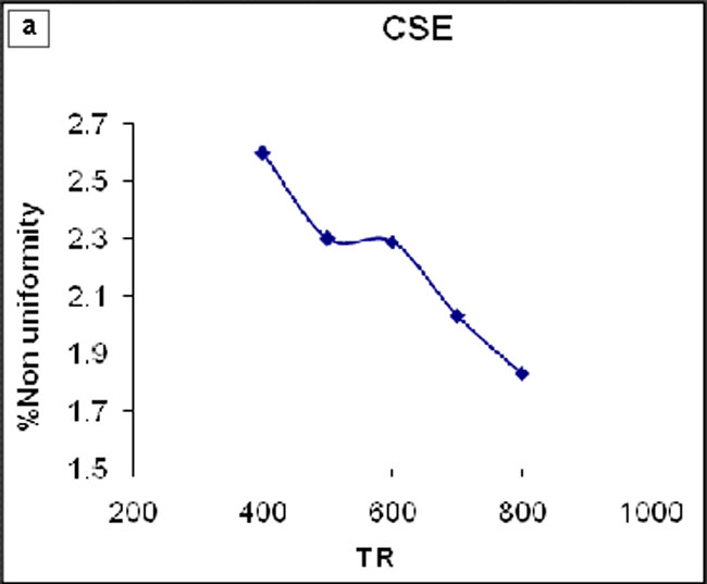
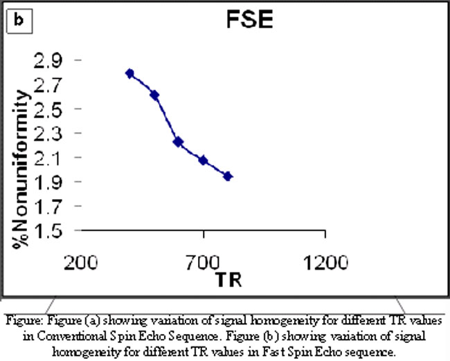
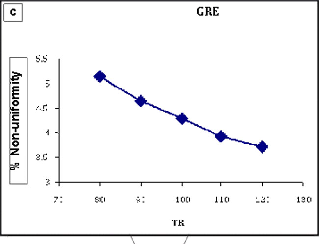
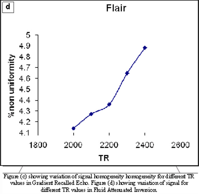
The Figures (a), (b), (c) and (d) show the variation of non-uniformity with different values of TR in different pulse sequences for T1 weighted images in FBX gel.
The average value of non-uniformity was calculated as 0.76%, 0.83%, 1.43% and 1.89% for CSE, FSE, GRE and FLAIR respectively.
Discussion
These results shows that signal non-uniformity decreases with the increase of repetition time for the first three sequences and that the results are unexpected for FLAIR. The use of relatively long TR times in FLAIR, influences in the sense that signal non uniformity increases. Another property of inversion-recovery sequences is linked to the choice of TI. If it is chosen such that the longitudinal magnetization of a tissue is null, the latter cannot emit a signal (absence of transverse and longitudinal magnetization), thus allowing the signal of a given tissue to be suppressed by selecting a TI adapted to the T1 of this tissue. The use of longer TI is chosen so that the signal from simple fluids, such as cerebrospinal fluid (CSF) is nulled. They have less signal intensity because the T1 contrast on inversion recovery images is based on variable rates of decay during the TI. All tissues lose signal intensity during this time but at different rates. The short TR used with this pulse sequence tends to reduce signal non uniformity. However, our findings of increased signal non uniformity with increasing TR may be related to the increased signal intensity of the gel phantom with the use of a relatively long TR, which generates a concordant increase in prominence of related non uniformity. Nonuniformity was defined as the percentage of the standard deviation of image signal intensity relative to its mean signal intensity.23,24 Variation of TE did not show any noticeable result for T1 weighted images in FBX gel.
Conclusion
This study demonstrates signal homogeneity for different pulse sequences with the change of MRI parameters in FBX gel. The probability of using this gel has been increased because of its better sensitivity and decreased diffusion rate. The non-uniformity of signal has been evaluated for CSE, FSE, GRE and FLAIR. Changes in TE did not show any predictable result for signal homogeneity. Effect of TR on CSE and FSE does not appear to be very critical. However, FLAIR is sensitive to TR changes. The choice of parameters is relatively easy for CSE and FSE, with image quality being maintained for different values of TR as compared to FLAIR. A careful selection is therefore, necessary with the use of FLAIR.
Acknowledgements
We are thankful to the Department of Medical Physics of Ninewells Hospital, University of Dundee, Scotland, UK for their great support and encouragement for this research work. We are also thankful to Higher Education Commission (HEC) of Pakistan for funding through its International Research Support Initiative Program.
References
1.Fricke H, Morse S. The chemical action of Roentgen rays on dilute ferrous sulphate solutions as a measure of dose. Am J Roent Radium Ther Nucl Med 1927; 18: 430-2.
2.Fricke H, E Hart. Chemical Dosimetry. In: Attix FH, Roesch WC. Editors, Radiation Dosimetry. Vol 2. New York: Academic Press, 1955.
3.Gore, JC, Kang YS, Schulz RJ. "Measurement of radiation dose distributions by nuclear magnetic resonance (NMR) imaging." Phys Med Biol 1984; 29: 1189-97.
4.Saur S, Strickert T, Wasboe E, Frengen J. "Fricke gel as a tool for dose distribution verification: optimization and characterization." Phys Med Biol 2005; 50: 5251-61.
5.De Deene Y. Gel dosimetry for the dose verification of intensity modulated radiotherapy treatments. Z Med Phys 2002; 12: 77-88.
6.Spinks, JWT, Woods RJ. Introduction to Radiation Chemistry. 3rd Ed. Toronto: John Wiley & Sons Inc, 1990.
7.John Raymond Greening. Fundamentals of Radiation Dosimetry 2nd Ed. Scotland: CRC Press, 1985.
8.Baldock C. Historical overview of the development of gel dosimetry: a personal perspective. Journal of Physics: Conference Series 56 (2006) 14-22.
9.Rae WID, Willemse CA, Lotter MG, Engelbrecht S, Swarts JC. Chelator effect on ion diffusion in ferrous-sulphate doped gelatin gel dosimeters as analyzed by MRI Med Phys 1996; 23: 15-23.
10.Noda S, Suzuki Y, Hoshino Y, Furukawa S, Katoh H, Kurotaki K, et al. Clinical application ofthe Fricke-Glucomannan gel dosimeter for high-dose-rate (192) Ir brachytherapy. Phys Med Hiol 2008; 53: 3985-93.
11.Luciani AM, Palma A, Adorante N, Barone P, Grande S, Ranghiasci C, et al. Optical images of dose distributions in Gel-Fricke: dosimetric performances of the gel. Radiat Prot Dosimetry 2006; 122: 455-6.
12.Appleby A, Leghrouz A. Imaging of radiation dose by visible color development inferrous-agarose-xylenol orange gels. Med Phys 1991; 18: 309-12.
13.Viti V, d\'Errico F, Pacilio M, Luciani AM, Palma A, Grande S, et al. Optical imaging of dose distributions in Fricke gels. Radiat Prot Dosimetry 2006; 120: 148-50.
14.Bero MA, Gilboy WB, Glover PM, El-masri HM. Tissue-equivalent gel for noninvasive spatial radiation dose measurements. Nucl Instrum Method B 2000; 166: 820-5.
15.Crescenti RA, Scheib SG, Schneider U, Gianolini S. Introducing gel dosimetry in a clinical environment: customization of polymer gel composition and magnetic resonance imaging parameters used for 3D dose verifications in radiosurgery and intensity modulated radiotherapy. Med Phys 2007; 34: 1286-97.
16.Afzal M, Lerski RA. Effect of the Time on Dose Response Sensitivity of Dosimeter Gels Between Preparation and Irradiation. JP & App Sc 2001; 20: 87-91.
17.M Afzal, Lerski RA. Temperature Dependence of Spin-Lattice Relaxation Times of Ferrous Sulfate Gels. JP & App Sc 2000; 19: 9-12.
18.Afzal M, Lerski RA. Time dependence of spin lattice relaxation time in Fricke gels. JP & App Sc 2000; 19: 25-9.
19.Afzal M, Lerski RA. Optimization of MR imaging sequence for the dosimeter gels of known T1 values. JP & App Sc 2000; 19: 39-47.
20.Afzal M, Lerski RA. Effect of concentration of ferrous ions on zero dose T1 value of dosimeter system. JP & App Sc 2000; 19: 71-3.
21.Kelly RG, Jordan KJ, Battista JJ. Optical CT reconstruction of 3D dose distributions using the ferrous-benzoic-xylenol (FBX) gel dosimeter. Med Phys 1998; 25: 1741-50.
22.Khan FM. Physics of radiation therapy. (Lippincott, Williams and Wilkins) USA: 2003.
23.Li T, Mirowitz SA. Fast T2-weighted MR imaging: impact of variation in pulse sequence parameters on image quality and artifacts. Magn Reson Imaging 2003; 21: 745-53.
24.Low RN, Francis IR, Sigeti JS, Foo TK. Abdominal MR imaging: comparison of T2-weighted fast and conventional spin-echo, and contrast-enhanced fast multiplanar spoiled gradient-recalled imaging. Radiology 1993; 186: 803-11.
Related Articles
Journal of the Pakistan Medical Association has agreed to receive and publish manuscripts in accordance with the principles of the following committees:


