
Kalimuddin Aziz ( National Institute of Cardio Vascular Diseases, Karachi. )
A. D. Memon ( National Institute of Cardio Vascular Diseases, Karachi. )
M. Rehman ( National Institute of Cardio Vascular Diseases, Karachi. )
A. Haque ( National Institute of Cardio Vascular Diseases, Karachi. )
August 1986, Volume 36, Issue 8
Original Article
RHEUMATIC FEVER AND CARDITIS AT THE NATIONAL INSTITUTE OF CARDIO-VASCULAR DISEASES-A FOLLOW UP STUDY
Abstract
Two hundred and forty nine children with rheumatic heart disease were seen at the National Institute of Cardlo Vascular Diseases (NICVD) during 1980-Oct. 1985. Seventy Seven per cent patients came from Kachi abadis of Karachi. Fifty four per cent had rheumatic fever with carditis, 38% acute reheumatic fever and 8% chronic rheumatic carditis. Mitral regurgitation was more frequently seen in the first two and mitral stenosis in the third group. Mortality was 6.8 % (JPMA 36: 196, 1986).
INTRODUCTION
Rheumatic fever and Rheumatic heart disease is most prevalent in poor communities in Pakistan. It is the commonest form of heart disease in Pakistani children. Studies by Ilyas showed an incidence of 11 per one thousand population in the rural communities of Sarhad1 (North west Frontier Province). Rheumatic heart disease was the predominant cause of admission for heart disease in a general children’s hospital2,3 and the incidence of patients with Rheumatic heart disease seen in Pediatric cardiac referal clinic of our institute was 9.4%.
It is true to say that comprehensive programmes of primary or secondary prevention of Rheumatic fever do not exist in Pakistan. There is also no general consensus among pediatricians and cardiologists as to the seriousness and magnitude of the problem. We are becoming more and more aware of the presence of coronary artery disease because the middle class professional is a sufferer from coronary artery disease and poor destitute, living in Kachi abadis of sprawling urban population in the sufferer from Rheumatic fever. Nation wide, Rheumatic fever and Rheumatic carditis is probably the most important form of serious heart disease which can be effectively prevented.
We present our cumulative experience with Rheumatic fever and Rheumatic heart disease in children who were seen and followed at the NICYD during 1980 to October 1985 period.
MATERIAL AND METHODS
Two hundred and forty nine children seen at the pediatric cardiology clinic from 1980 to Oct. 1985 were included in this study. The follow up visits were arranged according to the severity of heart disease at 3 monthly or shorter intervals. The hospital admission, special investigations such as Echocardiography and cardiac Catheterization, records were available for evaluation of severity of carditis. History of preceding sore throat and other manifestations of acute Rheumatic fever was obtained during clinical assessment; Electrocardiogram and chest X-ray examination were a part of assessment for all patients.
The information about death at NICYD was available. Eighty one patients who were only seen once were assumed lost to follow up and 168 patients were followed tifi 1985. The final assessment was made on the examination record of the day when patient was last seen and prior to October 1985. Therefore the follow up of each patient did not conclude till October 1985 but when the patient was last seen in the clinic.
The patients were refered to the paediatric cardiology clinic from all regions of Pakistan. The severity of heart disease was classified as mild, moderate or severe. The assessment was based on clinical examination, i.e. presence of respiratory distress, JYP elevation and liver size, cardiomegaly on plain chest x-ray film and presence of congestive cardiac failure and pulmonary edema or pulmonary Hypertension. Electrocardiogram and Echocardiogram were used to further define the severity of disease.
Patients with acute Rheumatic fever (ARF) were admitted in the hospital for 5-6 week period. For the first episode of ARF with moderately severe carditis and seriously sick children, prednisone was given in 1-2 mg/Kg daily dose for 1 week then reduced by half for the second week and reduced by¼ for the next 2-3 weeks and then gradually discontinued within 6 weeks. Prednisone was also given to all patients with pericarditis and congestive heart failure. Aspirin was not given concurrently with prednisone but added later when prednisone was being reduced. The dose of Aspirin was 75-100 mg/Kg/day in four doses administered after meals and was continued for 4-5 months or untill ESR had, returned to normal. All patients who did not require prednisone were given Aspirin at the initiation of treatment. Bed rest was advised for all patients with carditis. Ambulation was done when signs of congestive cardiac failure had been controlled with Digoxin, Frusemide and oral vasodilators such as Isosbide dinitrate. Other indicator for ambulation was the falling ESR and abatement of signs of acute activity.
Intramuscular long acting Penicillin such as penidure LA 1.2 and 0.6 million units, for greater than 60 pound weight and less than 60 pound Wt respectively, was given at admission and repeated at 2 weeks interval and then at 4 weekly intervals.
Desparately sick children with- congestive cardiac failure, vomiting or abdominal pain were given intravenous drugs such as Digoxin and Frusemide for cardiac failure and intravenous Hydrocortisone for Rheumatic activity. The intravenous therapy was continued till abdominal pain and vomiting subsided. Abdominal pain and vomiting was considered a serious complication and was noted in children who subsequently died due to acute Rheumatic activity.
RESULTS
Two hundred and forty nine patients were included in the study. The mean age at presentation was 9.7 ± 2.1 years (range 4-14 years). Eighty one patients were seen only once and 168 patients were followed for a mean period of 1.9 ± 1.5 years.
One hundred and fourteen patients required admission in the hospital; Sixty nine patients were admitted once, twenty three twice, and 22 more than three times. The diagnosis was made on history, clinical examination, Electrocardiogram and chest X-ray in 249 patients and confirmed by Echocardiography in seventy five (30%) and cardiac Catheterization in 12 patients (4.8%).
The mode of presentation was analysed for these 249 patients (Table I).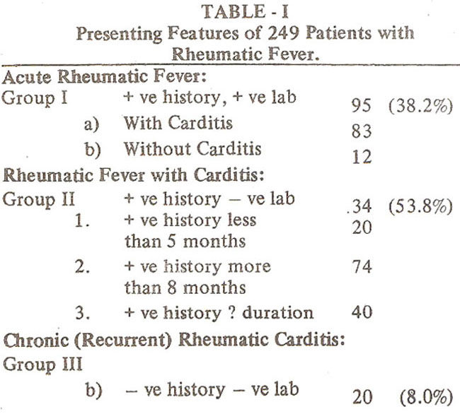
Acute Rheumatic fever was diagnosed in 95 patients by history of arthritis and or arthralgia severe enough to interfere with child’s locomotion, carditis, raised Antistreptolysin “0” titre and raised Erythrocyte sedimentation rate (positive laboratory). History of preceding throat infection was only occasionally elicited. Twelve patients in this group of 95 patients had no carditis (valvulitis) and eighty.
First episode of acute Rheumatic fever was noted in 69 of 83 patients and 26 of 83 patients had recurrent acute Rheumatic episode at the time of initial presentation. The distinction between first episode and recurrent Rheumatic fever was based on previous history of fever and Arthritis or presence of stenotic lesions when previous history was not available. Twenty of 69 patients(29%) with first episode of ARF presented in congestive cardiac failure. Mitral regurgitation was the most common single lesion and occurred in 47 of 69 patients. Mitral regurgitation and Aortic regurgitation was present in S patients and Isolated Aortic regurgitation in 4/one patient had congenital heart disease. Twelve of 69 patients with 1st attack of ARF presented with tachycardia, fever, arthralgia and positive laboratory tests but no evidence of valvulitis or cardiac murmur (Table II).
Four of 69 patients presented with chest pain due to pericarditis and pericardial effusion and also had evidence of mitral regurgitation.
Recurrent acute Rheumatic fever was diagnosed in 26 patients, 15 of these had mitral regurgitation, 6 had mitral and Aortic valve regurgitation and 4 had significant mitral stenosis and mitral regurgitation in 4 and one patient had Isolated Aortic valve regurgitation. Congestive cardiac failure was present in 6 of 26 patients (Table II).
The second group of 134 patients presented with evidence of carditis and a positive history of joint pains and fever but ESR & ASOT were within normal range at presentation (Table III).
Twenty of these patients presented with history of joint pains and fever of less than 5 months duration and 74 had history of more than 8 months duration. Forty one patients remembered episode of joint pain and fever but did not recall details or timing of the illness. The dominant lesion in this group was mitral regurgitation occurring in 84/134 (63%) patients, followed by mitral valve stenosis and mitral regurgitation and Isolated mitral stenosis in decreasing order of frequency. Isolated Aortic regurgitation was the least common lesion occurring in only 5 patients.
The third group of patients with Rheumatic carditis presented with evidence of valvulitis 12but no preceding history of joint pain or fever could be elicited and ESR & ASOT titre were within normal limits at the time of presentation. There were 20 patients in this group (Table IV).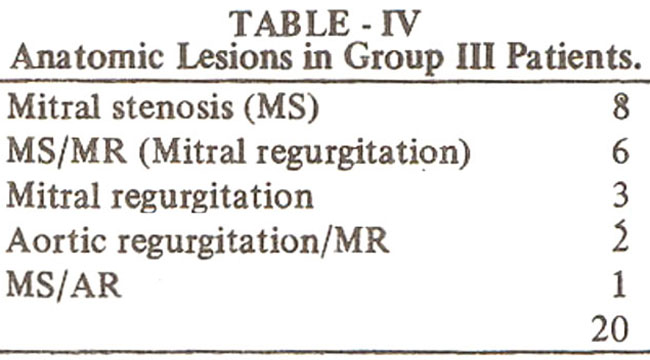
Mitral stenosis was the commonest lesion occurring in 8 patients followed by MS/MR in 6 of 20 patients.The spectrum of valve lesions and intra operative morphologic features of the stenotic mitral valves suggest that these were Rheumatic in etiology
Erythema margination and Rheumatic nodules or prolonged PR interval on the electrocardiogram were not observed in any patient. The diagnostic value of PR interval could not be assessed since detailed ECG reading was not performed routinely. Rheumatic chorea was observed in one patient who did not have signs of valvulitis.
The most frequent lesion among the groups was mitral Insufficiency which occurred in 59.8% of cases. Isolated mitral steñosis was noted in 8.4% and Isolated Aortic regurgitation was present in 4% (Table V).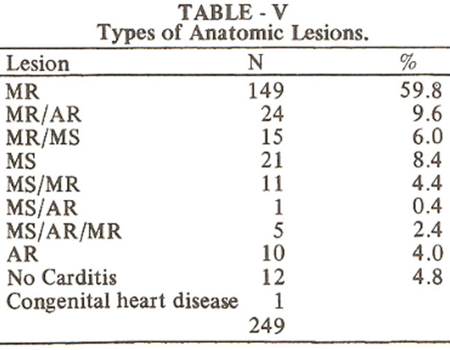
Tricuspid regurgitation was noted in 10 patients all of whom had severe pulmonary artery hypertension. Half of these patients had critical mitral stenosis. Bacterial endocarditis was noted in 8 patients, two of whom died and mitral regurgitation was the under lying lesion in six of these. Mitral stenosis alone or in combination with other valve lesions was present in 53 (2 1%) cases. The mean age of these was 9.9 ± 2.3 years range 7-14. In 21 patients Isolated mitral stenosis was present with, mean age of 9.5 age 10.6 ± 1 .8 years. In 16 patients mitral regurgitation was the dominant lesion in association with mitral stenosis with a mean age of 10.4 ± 2.1 years (Table VI).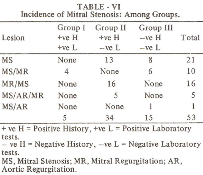
Geographic Distribution of Patients
Majority of patients (77%) came from Karachi (Sind) and only 15 patients (6%) were referred from Sind interior. Others iiicluded 3% from Sarhad, and 5% each from Punjab and Baluchistan. In 20 patients (8%) the address was not recorded. Analysis of data from Karachi referals showed that overwhelming majority of patients belonged to kachi Abadis. New Karachi, Malir, Landi and Korangi had the heaviest concentration of Rheumatic patients. The patients were however scattered through out the poor communities in the city (Table VII).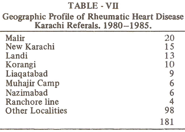
Presentation
The delay between the onset of fever and arthralgia and presentation at the NICVD could be determined in 188/249 patients. Only nineteen patients (10.1%) presented within 4 weeks of onset of symptoms and majority 118 of 188 patients (62.8%) presented between 4 weeks to 6 months, mean 2.6 ± 1.6 months. Twelve (6.4%) had a delay between 7-12 months and 39 patients (21%) presented after one year of the onset of ifiness.
The severity of cardiac lesion was evaluated at admission in 213 patients, mild degree of valvular damage was assessed in 48 (22.5%), moderate in 84 (39.5%) and severe in 81 patients (38%).
Follow Up
At admission congestive cardiac failure was noted in 65 of 249 patients (26%) and was still present in 29 of 168 (17%) patients on the final assessment. Recurrent Rheumatic fever was noted in 9 of 168 (5.3%); seventeen of 249 patients died, a mortality of 6.8%. Detailed analysis of the 17 deaths during the follow up period showed that 8 patients were seen only once, 5 patients were followed for less than 1 year and 4 patients were followed for more than 1 year.
Subacute bacterial endocarditis was noted in 8 of 249 patients during the follow up period, six of whom had mitral regurgitation. Two patients with bacterial endocarditis died. Causes of death on medical follow up were analysed and showed that majority (8 of 17) died due to cardiac failure consequent to Acute Rheumatic fever and majority of these had two or more valve lesions. Bacterial endocarditis (2 patients), pulmonary edema (2 patients), severe CHF (1 patient), cerebral embolism (1 patient) and Digoxin over dose (1 patient) accounted for the remaining deaths (Table VIII).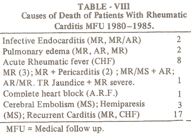
Assessment of the severity of disease was compared in 117 of 168 patients at first and final visit. Improvement was noted in only 18 (10.7%) patients and 64 patients (55%) remained unimproved and actual deterioration in the severity was noted in 35 (30%) patients. Of the 18 patients, who showed improvement on follow up, ten had congestive cardiac failure and 10 patients showed actual improvement in valve damage. Two patients lost the murmur of mild mitral regurgitation at 1.5 to 2.5 years of follow up.
Cardiothoracic ratio on chest X-ray film was available at 1st and last assessment in 43 patients and showed that CT ratio (0.61 ± 0.9) at the final assessment was greater compared to CT ratio (0.53 ± 0.08) at the initial assessment. Twenty four patients under went surgery with three immediate deaths (12.5%) and two late deaths (21%). Nine patients had mitral valvotomy, eight had valve replacement and 7 patients had mitral valve annuloplasty and repair. The overage follow up period of surgically treated patients was 2.9 ± 2.6 years.
DISCUSSION
Geographic Distribution
Our data, although referal based suggests that overwhelming majority of patients resided in the poor communities of Karachi city. The patients belonged to lower socioeconomic strata since majority lived in Kachi or semi developed communities. Other parameters of socioeconomic status as earning capacity, housing conditions, or family size was not determined and can be only indirectly infered. Significant delay from the onset of symptoms to the time of presentation at the NICVD suggests lack of parental understanding regarding the importance of fever and joint pains. The stage of tonsillitis is not even recalled because the mild illness is not considered significant for medical attention. This is an indication of how poor communities deal with minor infections and fever in their children. The medical help is not sought at the sore throat stage and in many instances it may not be easily available. Irregularity of clinic visits and lack of drug compliance are due to the lack of finances required for travel from home to the hospital. In many instances this incurs loss of day’s wages for the fathers. Lack of facilities for home help for mothers who would bring the child to the hospital for clinic visits is again a factor contributing to the irregularity of follow up visits.
Mode of Presentation
The delay in seeking medical advise, from the onset of disease i.e. fever and joint pains and presentation, poses a diagnostic problem because the features of Rheumatic fever and carditis have been modified by then and the raised ESR & ASOT may have returned to normal5. The history of preceding events and even the timing of the present event is not recalled with precision. The mode of presentations of acute Rheumatic fever is therefore variable as is suggested by various modes of presentation of our patients. Most of our patients did not present with fever and joint pains but with the symptoms resulting from carditis namely cardiac decompensation, tachycardia and shortness of breath.
A large number of patients in group II had acute rheumatic fever for more than eight months and some presented after few years of the event of acute Rheumatic fever. In this group the presentation is not of ARF but its sequelae namely carditis. In approximately a third of these no definite date of an event such as fever or joint pain or Tonsillitis could be elicited. All patients with carditis should have, throat culture, ESR & ASOT estimation so that activity can be detected, since in significant number of patients the dating of onset can-not be determined.
The third mode of presentation is of those children who present with delayed complications of carditis. The group is small, 20 out of 249 patients, and the common lesion in this group is mitral stenosis. Obviously a significant period must elapse between the onset of disease and development of mitral stenotic lesions. Although, ages can not be precisely determined in our patient population because recall of parents memory has to be relied upon for age determination, we have seen children as young as 7.-8 year with mitral stenosis who required mitral valvotomy. The question that may be raised is whether these steñotic mitral valves are congenitally malformed or have acquired Rheumatic affection. There are significant anatomic differences how ever, which suggests that these valves are affected by Rheumatic affection. The mitral valve annulus is normal and on Echocardiogram leaflet fusion can be observed, the leaflet size is not significantly reduced and leaflets are thickened at the edges and cordae tendinaè are short due to fibrotic process. The left ventricle is usually of an adequate size at surgery and papillary muscle abnormalities are not present. Close mitral valvotomy can be successfully performed. These valves have been observed intraoperatively in few cases and show features of commissural fusion and thicking. Further more in group I and II.
Patients with mitral stenosis, recurrent acute Rheumatic fever has been diagnosed by positive history and positive lab tests. rhis may not be a direct evidence but coupled with unusually high incidence of mitral stenosis in younger age provides a strong evidence for Rheumatic etiology. Only one patient with congenital cardiac malformation (LTGH & VSD) was seen who developed ARF diagnosed by a positive history and positive laboratory tests. Furthermore majority of patients (38/53) with mitral stenosis had a positive history and positive laboratory tests.
Medical Follow up
Comparision of the initial and final assessment of severity of cardiac lesions showed that only minority of patients showed improvement in the symptoms and even smaller number showed actual healing of lesions. The follow up period however may be top short to expect healing in significant number. The most discouraging aspect of the follow up results is that majority of patients either remained unimproved or actually deteriorated as far as the severity of lesions is concerned. The lack of improvement, recurrent episodes of cardiac decompensation and recurrent acute Rheumatic fever (5.3%) during the follow up period suggests non compliance with cardiac medications as well as penicillin prophylaxis. The usual practice in this group of patients with chronic disease is that once digitalis and anticongestive measures have improved symptoms, the drugs are discontinued. Our admission records show that nearly fifty percent of patients during the follow up period required two or more admissions for cardiac decompensation Or recurrence of Rheumatic fever. Admittedly some degree of the lack of improvement in the disease process may be related to the initial severity of cardiac damage since the valvular damage was assessed to be moderate to severe in degree in 75% of patients.
However socioeconomic constraints account, in large measure, for the irregular clinic visits, non compliance of maintenance drugs and penicillin prophylaxis. It seems to us that hospital based pilot programmes even if they can be laced with adequate personnel and drugs will not alter the natural course of Rheumatic carditis in our communities.
The present data shows that in our social setting once Rheumatic carditis has occurred the chance of relief or cure are small and therefore efforts at primary prevention should be initiated which may be undertaken in phases.
Primary prevention involves treatment of streptococcal tonsfflitis. Educational efforts in informing public of the danger of sore throat should be instituted and medical personnel should be made aware of treating sore throat difigently. Where throat culture facilities are not available, clinical tonsillitis and pharyngitis should be treated with 10 day course of oral penicillin. The medication is cheap and safe. One may be over treating viral tonsillitis but cases of streptococcal tonsillitis will not be missed.
The improvement in socioeconomic conditions should be, the final goal of any society. These are long term goals but in the interim treatment of tonsillitis or pharyngitis should be undertaken seriously. Parents, physicians and paramedical personnel should be made aware of this need through communication media.
Role of Surgery
There are few options as far as surgical procedures are concerned. Simplest of these is close mitral valvotomy. It is the most economical and effective means of relieving symptoms and improving excercise capacity in our geographic setting. It however leaves, the valve in damaged state and prone to recurrent Rheumatic activity and bacterial endocarditis and restenosis. Mitral valvotomy has borne the best results in the present study. However its place in management of Rheumatic valvular disease in children is limited since only 8.4% of our patients had pure mitral stenosis.
The other two surgical options require open heart procedure and cardiopulmonary by pass. Commonly mitral regurgitation is the lesion which requires surgery. In our series more than half of children had isolated mitral insufficiency. There are 2 surgical options to deal with regurgitant mitral valve. The first option is to repair the valve and perform annuloplasty that is narrowing of the valve annulus. The advantage of this procedure is that patients own valve is left in place and post operative anticoagulants are not required. Our preliminary results show that significant symptomatic relief can be obtained although hemodynamics are not completely restored to normality. Although in few children we have observed late deterioration and increasing cardiomegaly, we however believe that increasing surgical expertise in this regard is bound to lead to more fruitful outcome and that valve repair is a feasible option in our Socio-economic setting. The second surgical option is to replace the valve with tissue valve or metal valve disc or ball type prosthesis. The medical constraints placed on the patient after the valve replacement are too great for these group of patients.
There is the burden of anticoagulant therapy and repeated prothrombin time estimation and variation of the dose of anticoagulants, repeated visits to ensure the functioning and viability of the valve, which requires a specialised hospital not in reach of the patients in farther areas of our villages. We do not believe that even though near normal hemodynamics can be achieved by the valve replacement that it is of a lasting value and economically and logestically a sound form of therapy in our socioeconomic setting.
Our data shows. that careful medical management results in identical mortality when compared to surgery. Therefore Rheumatic carditis should be managed medically whenever possible because the surgical options are not entirely satisfactory in our geography.
It is concluded that the degree of carditis is severe with significant morbidity and mortality. Medical and surgical management of these patients has not produced any significant improvement or cure of valvulitis. Recurrent Rheumatic fever and recurrent cardiac décompensation episode are due to noncompliance of drugs.
The prognosis of a child developing Rheumatic carditis in our geographic setting remains guarded.
REFERENCES
1. Ilyas, M., Peracha, M.A., Ahmed, R., Khan, N., All, N. and .Janjaua, M. Prevalence and pattern of rheumatic heart disease in the Frontier Province of Pakistan. JPMA., 1979; 29: 165.
2. Rahimtoola, R.J., Shafqat, H. and Ramzan, A. Acute rheumatic fever and rheumatic carditis in children. Pakistan Heart J., 1980; 13: 2.
3. Rahimtoola, R.J., Majid, I., Shafqat, H. and Qureshi, A.F. Congenital heart disease in children visiting J.P.M.C. Pakistan Heart J., 1980; 13:21.
4. Aziz, K. Incidence of heart disease in children at the National Institute of Cardiovascular Diseases. JPMA., 1984;34:300.
5. Padmavati, S. Epidemiology of cardiovascular disease in India. 1. Rheumatic heart disease. Circulation, 1962; 25 : 703.
Journal of the Pakistan Medical Association has agreed to receive and publish manuscripts in accordance with the principles of the following committees: