
Muhammad Zahoor Janjua ( Department of Anatomy, Jinnah Postgraduate Medical Centre, Karachi. )
Umar Draz ( Department of Anatomy, Ayub Medical College, Abottabad. )
May 1991, Volume 41, Issue 5
Original Article
ABSTRACT
The effect of paracetamol against ulcerogenic agent, naprosyn on the gastric mucosa of albino rat was observed under dissecting as well as laboratory microscope. Paracetamol in a dose of 250 mg/kg body weight provided protection against the ulcerogenic effect of naprosyn under dissecting microscope. Under laboratory microscope, a significant increase in the mucosal thickness with the administration of paracetamol followed by naprosyn was observed, while the flattening of the surface epithelium with slight exfoliation may be attributed to relative increase of pepsin from the chief cell. The increased secretory activity of the mucous neck cells in animals treated with paracetamol followed by naprosyn may be due to the increased bio-synthesis of prostaglandin from these cells which might have produced protective influence against the damaging effect of naprosyn(JPMA 41: 107, 1991).
INTRODUCTION
Several non-steroidal anti-inflammatory drugs (NSAID) are believed to act by inhibiting the biosynthesis of prostaglandm from arachidonic acid since it is presumed that prostaglandin acting on the brain may cause fever. This may explain why certain anti-inflammatory agents are also anti-pyretic. The anti-pyretic action of paracetamolis no doubt much more effective in preventing prostaglandin formation in brain than in tissues3. In rats, paracetamol given intragastrically thirty minutes before the administration of absolute ethanol or acidified aspirin dose dependently, reduces the formation of mucosal lesions4. One would presume that paracetamol would also protect the stomach against the ulcerogenic effect of other NSAID. To test this hypothesis, naprosyn (naproxin sodium) commonly used in the treatment of rheumatoid arthritis and other rheumatic or musculoskeletal disorders, dysmenorrhoea and acute gout for its antipyretic, anti-inflammatory and analgesic actions was selected for the present study. Its ulcerogenic effects result in gastric discomfort, dyspepsia, heart burn, nausea, vomiting and gastric bleeding.
MATERIALS AND METHODS
Adult male albino rats weighing between 200 and 350 grams, bred in animal house of Jinnah Postgraduate Medical Centre, Karachi were used in the present study. Naprosyn (Alkaloid, Hungary) and paracetamol were administered as suspension in one percent gum acacia in distilled water by oral intubation in a volume of one ml per 100 gin body weight. The ulcerogenic dose of naprosyn used was 7.1 mg per kg body weight2. The protected dose of paracetamol was assessed by administering different doses (ranging between 30-250 mg/kg body weight) against naprosyn. The dose of 250 mg/kg body weight was found optimum to protect mucosal lining of the stomach and was used for further detailed study. A total of 40 animals used in present study were divided into four groups; A,B,C & D, each group had 10 animals. The animals from each group were kept on fasting for 24 hours; water, however, was available to them freely. The animals were sacrificed 6 hours after the treatment.
Group A
Served as control, were given gum acacia in the same volume as used in treated groups.
Group B
Received naprosyn only.
Group C
Received gum-acacia in same volume as used for paracetainol followed by naprosyn.
Group D
Received paracetamol followed by naprosyn. The erosion produced by naprosyn was observed under dissecting microscope and assessed according to the procedure adopted by van Kolfschoten et al. 1983 and score was given to the erosion according to arbitrary scale of Bonta1 (Table I). The cumulative score of one group was divided by the number of animals and expressed as median erosion score of the group. These erosions were subjected to detailed histological study with the help of laboratory microscope to observe the change in the thickness of mucosa, and the distribution and height of the mucous, mucous neck, chief and surface epithelium, parietal cells of the gastric gland to assess their secretory activity. The general morphology of the erosion was studied under 6 micron thick paraffin embedded H&E stained sections and the count of secretory cells in PAS stained sections. The cells count was done under 40X objective & 8X ocular in a strip covering the whole field measuring 150Mm in width extending from the surface to the base of gastric gland.
Statistical analysis of the data.
Statistical evaluation was done using student’s test. The difference was regarded statistically significant if the ‘P’ value was equal to or less than 0.05.
OBSERVATIONS AND RESULTS
A) Normal control group:
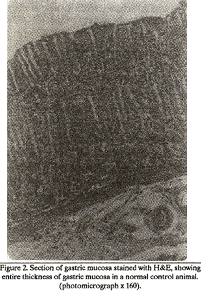
Opening along the greater curvature, the stomach in most of the (8) animals revealed food contents mixed with pale yellow fluid. However, in the remaining two animals little muddy coloured food contents were observed. Under dissecting microscope the internal surface of each stomach was clearly identified into two parts byaraised ridge. The greyish white part continuous with oesophagus was squamous part (rumen) while the pink area raised into folds (rugae) continuous with duodenum was identified as secretory or glandular part Under laboratory microscope, H&E stained sections of the stomach revealed all the layers; mucosa, submucosa, musclaris and serosa. The thickness of the mucosa observed was 532 ,um + 29.77. The surface epitheium composed of mucous secreting tall columnar cells with eosinophilic granular cytoplasm and basal location of round or oval nuclei. The surface mucous cell count was 51.20 + 3.52with the mean height of 12.48 + 0.72 1um. The gastric glands round to tubular in shape were lined by three types of cells; mucous neck, chief and parietal cells. The mean height of mucous neck cells observed was 8.88 um + 0.30. These cells secrete mucous, showed pale vacuolated cytoplasm with basal nuclei perpendicular to the long axes of the cells and their mean cell count was 29.14 + 3.09 cell/unit area. The mean height of the pyramidal shaped chief cells observedwas 8.20Mm + 0.38 havinground or oval nuclei showing basophilic granular cytoplasm and apical vacuolations, were located at the base of the gland. The mean values of the chief cells count was 81.2 + 6.89/unit area. The large ovoid parietal cells, with light pink to eosinophilic cytoplasm and central nuclei, lie at adluminal side between chief cells and basement membrane at the base of the gland. The mean cell count observed was 54 + 3.97 /unit area while mean size of these cells was 12.5 um + 0.26 (Figure 2).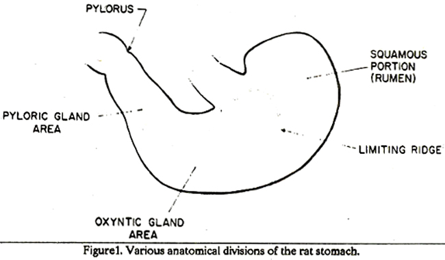
B) Naprosyn treated group:
Opening the stomach along the greater curvature, 3-5 dark brown patches were observed in fundic area which appeared circumscribed elongated erosions under dissecting microscope (Figure 3).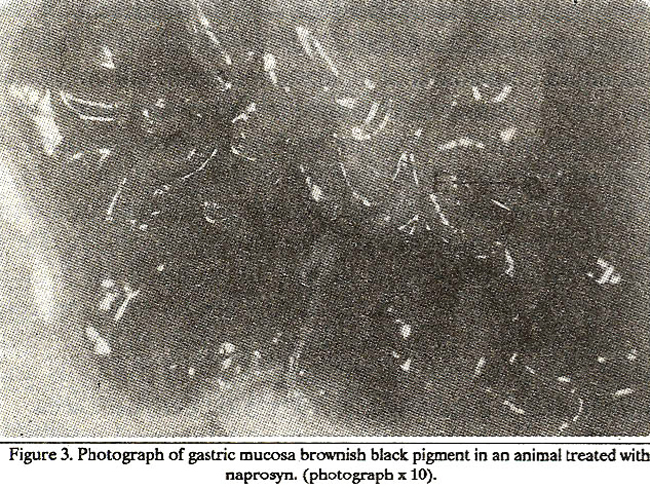
The median erosion score was calculated as 4.6 + 0.79. Numerous spider web like tortuous dilated blood vessels were seen on rugaes of the stomach. Under light microscope, the mean mucosal thickness was 386.4 + 14.8 pm which showed statistically significant decrease when compared with group A & D (Table II) and exfoliation of the epithelial cells with moderate degree of pyknosis of nuclei and dilatation of blood vessels were the common features. A circumscribed dark brown erosion area covering 3/5 to 4/5 of the depth of the mucosa inifitrated the lamina propria between the glands causing, necrosis of the epithcium as well as the tubules of the gland, however, numerous partial cells with pyknotic nuclei were observed. Lymphocytes, plasma cells and neutrophils were observed within and around the erosions (Figure 4).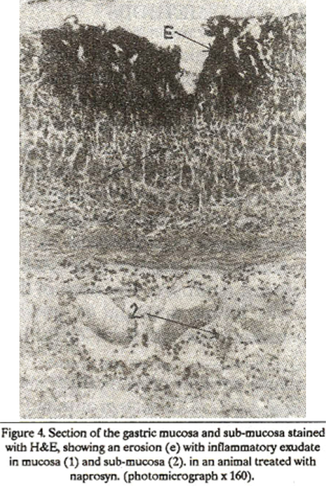
A significant decrease in the surface mucous cell count (27.13 + 1.82) was seen when compared with group A& D (Table 1).
The mean height of these cells (8.9 pm + 0.37) showed significant decreased values when compared with group A, C & D (Table III). A significant decrease in mucous neck cells count (18.6 + 0.60) was noticed when compared with group A & D (Table II). and the height of these cells (9.5 pm +0.37) decreased as compared to group D (Table III). The mean values of the parietal cell count (59.26 + 3.54) decreased significantly when compared with group C(Table II) while the size of these cells (11.72pm + 0.17) decreased when compared with group A (Table III). The mean value of the chief cells count (55.19 + 0.80) was observed to decrease significantly when compared with group A, C and D (Table II),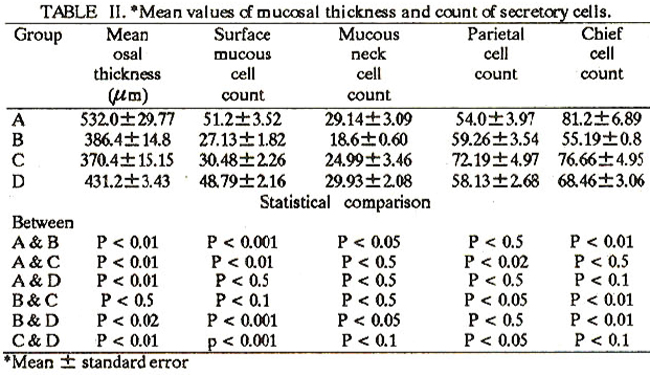
whereas the mean height of these cells (8.88Mm + 0.24) was relatively increased when compared with group A & C and decreased when compared with group D, but was not statistically significant (Table III).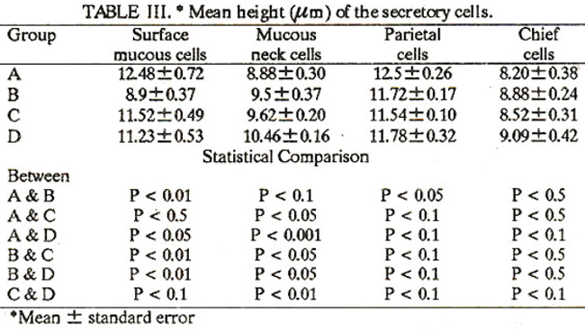
C) Gum Acacla followed by naprosyn treated group
Similar findings were observed in this group as in naprosyn treated group B with regard to the type of erosions and histological features but the erosions score and the inflammatory exudate around the erosion decreased in intensity (Figure 5).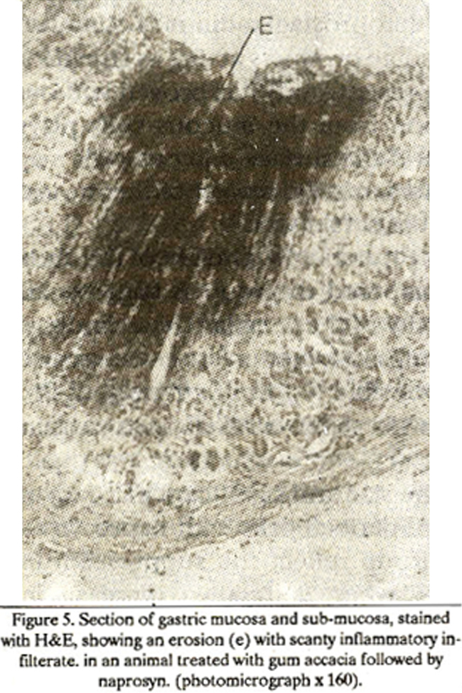
The median erosion score (3.80 0.37) decreased when compared with group B but was not significant statistically. The mean mucosal thickness (370.4 um + 15.15) decrease significantly when compared with group A & D (Table II). The mean values of the surface mucous cells count (30.48 + 2.26) was observed to decrease significantly when compared with group A & D (Table II), while the mean height of these cells (11.52um + 0.49) increased significantly when compared with group B (Table III). The mean values of the mucous neck cell count (24.99 + 3.46) showed no significant change when compared with group A, B and D (Table II). whereas the mean height of these cells (9.62 Mm + 0.20) decreased significantly when compared with group D (Table III). The mean values of the parietal cell count (72.19 + 4.97) increased significantly when compared with groups A & D (Table II) while the mean size of these cells (11.54 um + 0.10) decreased relatively when compared with groups A, B & D but was not statistically significant (Table III). The mean values of the chief cells count (76.66 + 4.95) increased significantly when compared with group B (Table II) while the height of these cell (8.52um + 0.31) was relatively increased when compared with group A and decreased when compared with groups B & D but was not significant statistically(Table III).
D) Paracetamol followed by naprosyn treated group
On examination, the mucosal surface of the stomach was pink coloured in the fundic and antral area and greyish white in the rumen area. Few spider like dilated blood vessels were observed over the convexities of the rugae on the anterior and posterior surfaces of the fundic area near greater curvature. Under laboratory microscope, mucosa showed normal histological appearance except slight dilatation of blood vessels and flattening of epithelial cells with pyknotic nuclei in certain areas (Figure 6).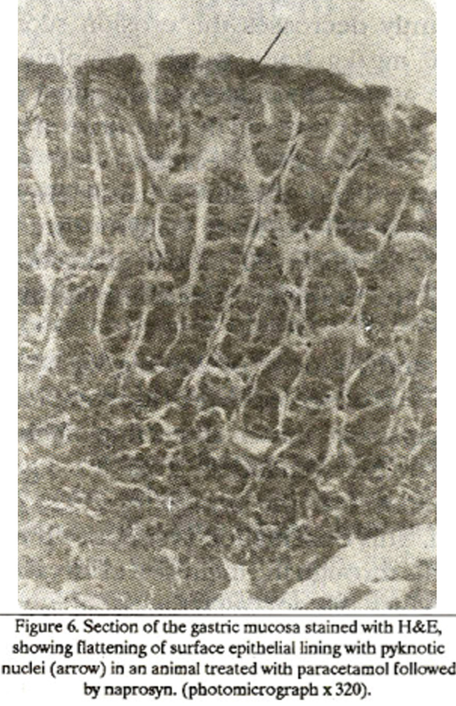
The mean mucosal thickness (431.2Mm + 3.43) decreased significantly when compared with group A while in creased significantly when compared with group B & C respectively (Table II). The mean values of the surface mucous cell count (48.79 + 2.16) increased significantly when compared with group B & C (Table 1), while the mean height of these cells (11.23 um + 0.53) increased significantly when compared with group B (Table III). The mean values of the mucous neck cells count (29.93 + 2.08) increased significantly when compared with group B (Table II), while the mean height of these cell (10.46 Mm + 0.16) increased significantly when compared with groups A, B & C (Table III). The mean values of the parietal cells count (58.13 + 2.68) decreased significantly when compared with group C (Table II), while the mean size of these cells (ll.78Mm + 0.32) showed no significant change when compared with groups A, B & C (Table III). The mean values of the chief cells count (68.46 + 3.06) increased significantly when compared with group B (Table II), while the mean height of these cells (9.09Mm + 0.42) increased relatively when compared with group A, B and C but was not significant statistically (Table III).
DISCUSSION
The study was designed to observe the protective effect of paracetamol against ulcerogenic agent. Naprosyn by localizing the site of lesion under dissecting microscope, and there after subjecting the sites to the detailed morphological study with the help of laboratory microscope for changes in the thickness of the mucosa, the epithelium and gastric glands with reference to their various secretory cells. As regard the protective dose of paracetamol, the data available showed a wide range of variation presented by different investigators. One group of workers6 reported that 30 mg/kg body weight of paracetamol significantly decreases the erosion score and the dose of 150 mg/kg body weight completely protects the stomach against the ulcerogenic effect of aspirin while another group observed that the dose of8O mg/kg body weight significantly decreases the ulcerogenic affect of aspirin and absolute ethanol. However, a third group9 used 500 mg/body weight to study the protective effect and observed partial protection against indomethacin and complete protection against ethanol (66% v/v). In the present study, a dose of 250 mg/kg body weight of paracetamol was observed to provide complete protection against naprosyn under dissecting microscope; however it can be concluded from the light microscopic obsenrations that the protective effect of paracetamol is partial. This observation is supported by the experimental results through the inclusion of an extra group-C in our study which did not catch the attention of the previous workers. The mechanism of protection can be explained by the hypothesis put forward by investigators9 who reported that paracetamol can stimulate the bio-synthesis of prostaglandin which may probably be responsible for this protective effect, although earlier workers5 reported similar results as they observed protection of gastric mucosa by prostaglandin against absolute ethanol, 0.6 N hydrochloric acid, 0.2 N sodium hydroxide, 25% sodium chloride and even boiling water. However, few workers4,7 reported that no protection was observed when administered indomethacin (a prostaglandin inhibitor) before the administration of paracetamol against ulcerogenic agents. In the present study the mucous contents of the stomach in group D (paracetamol followed by ulcerogenic agent) showed moderate increase as compared with group B and C observed by PAS staining under light microscope. This is in agreement with previous studies8 which were based on the culture of fundic epithelial cells, showed increased mucous by prostaglan din E2 in the gastric mucosa of the rat. The number and height of the surface mucous cells per unit area showed significant decrease in group B and C when compared with the normal controls; however, a significant increase in group D as compared to group B revealed that there has been recovery in the secretory activity of the cells under paracetamol treatment. The decreased height and number of mucous neck cells with naprosyn reflect decreased activity while increase in the number and height of these cell in group D as compared to group A and B show elevated secretory activity of mucous by the mucous neck cells which might have provided a protective shield or barrier between damaging agent and the surface mueosa. Our observations are in conformity with the previous authors5 but their findings were deficient and only ambiguous suggestion was made stating that prostaglandin may stimulate gastric cells to secrete some products acting as shield for mucosa, whereas, the present study has proved without any doubt that secretion from the mucous cells plays major protective role and the surface mucous cells may be minor contributor towards the revival of the normal activity under the effect of paracetamol. The increase in the number and height of the chief cells in group D as compared to group B suggests an elevated secretory activity of these cells. These findings reveal that increased pepsin secretion from the gastric mucosa might be responsible for the flattening of the surface mucous cells with slight exfoliation at certain places in group D. The decrease in the size and significant increase in number per unit area of parietal cells with naprosyn as compared to normal group reflect the suppression in activity of these cells, whereas relative increase in number without affecting the size of the cells in group D was observed when compared with group B. This is in contradiction to the usual belief that the increased parietal cell activity has damaging effect on the gastric mucosa. However, our findings could be correlated with some workers5 who stated that the protection of gastric mucosa is neither by decreasing the gastric secretion nor by the amount of hydrochloric acid. The mucosal thickness decreased relatively in group D when compared with group A, whereas increased significantly when compared with group B and C which shows partial protection by paracetamol. In the light of the above facts and figures, we can say that paraeetamol provides partial protective effect to gastric mucosa against naprosyn in the dose of 250 mg/kg body weight. The increase dosage may provide complete protection but with a risk of hepatotoxicity and nephrotoxicity. Therefore, this protective dose of paracetamol can be used to cover the risk of ulcerogenic drug administration with empty stomach. This study may act as a base-line for the extension of the project to the humans which may bring fruitful results in minimizing the danger of erosions when ulcerogenic drugs are used in combination with paracetamol in patients. The variations in the effect of these ulcerogenic agents used in the present study can be presumed to be either due to difference in the cellular response or their mode of action on different types of cells.
REFERENCES
1. Boots, L LA study of the effect of some glucorticoids and ACTH on artificially induced gastric ulcers of the rat. Arch. Intern. Pharmacodyn., 1961; 132: 147.
2. Diarnantis, W., Melton., .1., Sofia, R.D. and Ciofalo, V.B. Comparative gastric ulcerogenic effects of meseclazone, 5- Chlorosalicylic acid and other nonsteroidal anti-inflammatory drugs following acute and repeated oral administration to rats Toxicol. App!. Pharmacsl., 1980;52:454.
3. Girdwood, R.H. Dilling's clinical pharmacology. 25th ed. London, Bailliere Tindall, 1984,p.107.
4. Konturek, Si., Brzozowski, T., Piastucki, I. and R.adecki, T. Prevention of ethanol and aspirin-induced gastric mucosal lesion by paracetamol and salicylate in rats; role of endogenous prostaglandin. Gut, 1982; 23: 536.
5. Robert, A., Negamis, i.E., Lancaster, C. and Hanchar, A.J. Cytoprotection byprostaglandins in rats; prevention of gastric necrosis produced by alcohol, HCl, Na OH, hypertonic NaCI and thermal injury. Gastroenterology, 1979; 77: 433.
6. Seegers, A. J., Jager, LP., Van Noordwijk, J. Effect of phenacetin, paracetsmol and caffeine on theerosive activityof add in the rat stomacb; dose.response relationship, time course of erosion development and effects on acid secretion. J. Pharin. Pharmacol., 1979;31: 840.
7. Stem, A.I., Hogan, D.L, Kahn, LH. and Isenberg. J.I Protective effect of acetarninophen against aspirin and ethanol- induced damage to the human gastric mucosa. Gastroenterology, 1984;86:728.
8. Terano, A., lye K.J., Stachure, J., Sekhon, S., Hosajima, H., Mckenzie, W.N.Jr., Krause, W.J. and Wyche, J.H. Cell culture of rat gastric fundic mucosa, Gastroenterology 1982; 83: 1280.
9. Van Kolfschoten, A. A., Zandberg. P., Jager, L P., Noordwijk, 3. V. Protection by paracetamol against various gastric irritants in the rat. Toxicol. AppL Pharmacol., 1983; 69: 37.
Journal of the Pakistan Medical Association has agreed to receive and publish manuscripts in accordance with the principles of the following committees: