

Anjum Rehman ( Department of Nephrourology, Jinnah Postgraduate Medical Centre, Karachi-35. )
S.A.J. Naqvi ( Department of Nephrourology, Jinnah Postgraduate Medical Centre, Karachi-35. )
November 1980, Volume 30, Issue 11
Original Article

Abstract
Serum and urinary uric acid were estimated in two hundred apparently healthy males and females of different age groups, representing various socio-economic status.
The maximum levels of mean serum uric acid were found in the age group of 30-39 years in both sexes whereas mean levels of urinary uric acid in males were highest in the age group of 20-29 and in females between 40-49 years (JPMA 30:242 1980).
Introduction
Uric acid was isolated from a urinary concretion and was found to play a significant role in the evolution and ecology of vertebrate life (seegmiller et al., 1963). It is a nitrogenous waste product of aminoacid metabolism by way of purines (Gutman and Yu, 1965). Uric acid nitrogen constitute 60-90% or more of the total urinary nitrogen (Benedict et al., 1949).
Urate appears in blood mainly in the free form and the serum urate concentration represents a dynamic balance between uric acid production and disposition (Seegmiller et al., 1963). Uric acid production can be viewed in terms of denovo synthesis, reutilization and degradation (Thier and Wessler, 1974). Production may be regulated by the amount of various substrates which include ribose 5 phosphate, glutamine, glycine and aspartic acid and their induction of increased activities and amounts of enzyme involved in uric acid synthesis (Muramatsu and Kumura, 1979). Serum uric acid may be considerably increased in starvation, this is due to accelerated tissue turnover and to decreased renal excretion of uric acid probably resulting from acidosis that accompanies starvation (Toad, 1969). In normal human subjects renal excretion accounts for 57 77 percent of the body\'s daily production of uric acid (Seegmiller et al., 1961). Most of the remainder is excreted into gastrointestinal tract where it is degraded to allantoin, allantoic acid and other products by enteric bacteria (Sorenson, 1960). A remarkable feature of the renal regulation of uric acid excretion in normal man is the low uric acid clearance (Gutman and Yu, 1957). Impaired renal function decreases the uric acid excretion in the urine (Seegmiller et al., 1961).Uric acid determinations were undertaken in order to define the distribution characteristic of uric acid levels in normal subjects and to evaluate the importance of age and sex as variables in relation to the uric acid level.
Material and Method
Two hundred blood and twenty four hour urine samples from apparently healthy individuals of both sexes were collected. The age and sex distribution is shown is table 1.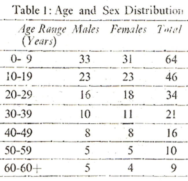
Serum and urinary uric acid were estimated by alkaline phosphotungstate assay of Caroll et al (1971).
Residts
In the present study the mean serum uric acid concentration for two hundred subjects of both sexes was 3.9 mg% and the range was 2.0-7.0 mg%. The mean value of all male subjects was 3.98 mg% and the range was 2.0-7.0 mg%. In females the mean value was 3.86 mg% and the range was 2.0-6.4 mg%.
The mean urinary uric acid concentration in two hundred apparently healthy males and females was 0.46 gm/24 hour and the range was 0.12-0.7 mg/24 hr. In males the mean value was 0.47 gm/24 hr and the range was 0.19-0.7 gm/24 hr and that for female the mean value was0.45 gm/24 hr and the range was 0.12-0.7 gm/24 hr.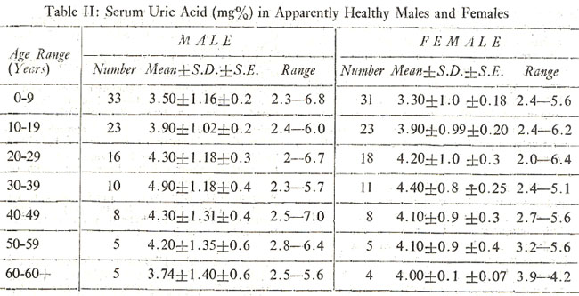
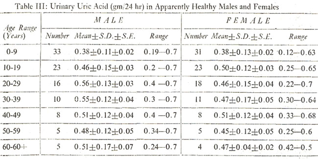
Table II and III represent serum and urinary uric acid concentration in apparently healthy males and females of various age groups.
Discussion
Uric acid serves as an important end product of purine metabolism. The metabolically important purines, adenine and guanine serve as building blocks for nucleic acid and as components of cofactors for a wide variety of biochemical reactions. Adenine and guanine originate either from purines or from a denovo biosynthesis. The purines formed in excess of the body\'s requirements as well as a portion of those arising from tissue catabolism undergo an irreversible oxidation to uric acid. Purine biosynthesis has been shown to be under the control of feed back mechanism that serves to regulate the rate of purine biosynthesis (Mclall and Magasanik, 1960).
A trend for increased mean uric acid value was observed with the advancing age and with a difference in values between the sexes Todd, 1969). This is in accordance with the present study. The data presented in Table II indicates clearly that age as well as sex bears a definite relationship to serum uric acid concentration. The values are low in children than those for the adult age range. During the age range 20-39 years the means in female subject remain approximately unchanged. It was found that mean serum uric acid level was slightly higher in males than females in all age groups except in sixth decade where serum uric acid in female was greater than male. These age sex trends generally confirm the experience of previous investigators. The findings of low serum uric acid levels in children without much differences between the sexes was reported by Wolfson et al (1949) and Mikkelsen et al (1965). Previous studies of Smyth (1957) showed a slight rise with the increasing age. This is also in accordance with the present study. Although not entirely in agreement previous study of Hauge and Harvald (1955) found no evidence of an age effect in women aged 30-79 years. In this series mean serum uric acid in female subjects remained relatively constant in the age group 40-60+ years.
Mikkelsen et al (1965) reported that age and sex relationship strongly suggest endocrine influences on serum uric acid concentration. This statement holds true in this study. Androgens may be important regulatory factors is suggested in the study by the following findings. Prior to puberty values are more or less similar in male and female subjects. In males values show a rise at the time of puberty and exceed those of female subjects during the age range 20-39 years. Estrogen and possibly progesterone are also of importance is indicated by the demonstration that female subjects have a lower uric acid level. It is suggested that estrogens may have a urate depressing ability.
In case of urinary uric acid, it was found that in males there was a gradual increase in the urinary excretion upto the age of 20-29 years, after that an increase in age was not followed by a subsequent increase or decrease of urinary uric acid and the values were more or less in the same range. In female, children of 0-9 years showed lower levels as compared to adults and the urinary excretion of uric acid was highest in the age group of 40-49 years (Table III).
References
1. Benedict, J.D., Forsham, P.H. and Statten, D. Jr. (1949) The metabolism of uric acid in the normal and gouty human studied with the aid of isotoic uric acid. J. Biol. Chem., 181:183.
2. Caroll, J.J., Coburn, H., Douglass, R. and Babson, A.L. (1971) Clin. Chem., 17:158.
3. Gutman, A.B. and Yu, T.F. (1957) Renal function in gout; with commentary on renal regulation of urate excretion and the role of the kidnev in the pathogenesis of gout. Am. J. Med., 23:600.
4. Gutman, A.B. and Yu, T.F. (1965) Uric acid metabolism in normal man and in primary gout. N. Engl. J. Med., 273:252, 313.
5. Hauge, M. and Harvald, B. (1955) Heridity in gout and hyperuricaemia. Acta Med., Scandinav. 152:247.
6. Mcfall, E. and Magasanik, B. (1960) Control of purine biosynthesis in cultured mammalian cell. J. Biol. Chem, 235:2103.
7. Mikkelsen, W.M., Dodge, H.J. and Valkenburg, H. (1965) The distribution of serum uric acid values in a population unselected as to gout or hyperuricaemia. Am. J. Med., 39:242.
8. Muramatsu, T. and Okumura, J. (1979) Effect of dietary methionine and arginine on uric acid excretion of cocks fed a protein-free diet, J. Nutr., 190:1057,
9. Seegmiller, J.E., Grayzel, A.I., Laster, I., and Liddle, I. (1961) Uric acid production in gout. J. Clin. Invest.,40:1304.
10. Seegmiller, J.E., Laster, L. and Howell, R.R. (1963) Biochemistry of uric acid and its relation to gout. N. Engl. J. Med., 268:712.
11. Smyth, C.J. (1957) Heriditary factors in gout; a review of recent literature. Metabolism, 6:218.
12. Sorenson, L.B. (1960) The elimination (of uric acid in man studied by means of C14-labeled uric acid, Uricolysis. Scand. J. Clin. Lab. Invest., 12, (Suppl. 54):1.
13. Thier, S.O. and Wessler, S. (1974) An approach to disorders of uric acid metabolism. Arch. Intern. Med., 134:579.
14. Todd, J.C. Clinical diagnosis by laboratory method, edited by Israel Davidson and John Bernord Henry. 14th ed. Philadelphia, Saunders, 1969.
15. Wolfson, W.Q., Krevsky, D., Levine, R., Kadota, K. and Cohn, C. (1949) Endocrine factors in gout; the significance of differences in childhood and adult urate metabolism. J. Clin. Endocrin., 9:666.
Journal of the Pakistan Medical Association has agreed to receive and publish manuscripts in accordance with the principles of the following committees:


