

Nayyar A. Masudi ( Institute of Physiology, The University, Glasgow G12 800, Scotland, U.K. Present Address: Physiology Department, Chandka Medical College Larkana, Sind. )
Desmond P. Gilmore ( Institute of Physiology The University, Glasgcow G12 8QQ, Scotland U.K. )
Catherine A. Wilson ( Department of Obstet. & Gynaecology, St. George’s Hospital Medical School. University of London, Cranmer Terrace, London, SW17 ORE. )
Alan S. McNeilly ( MRC Unit of Reproductive Biology, Centre for Reproductive Biology, 37 Chalmers Street, Edinburgh EH3 9EW. )
November 1985, Volume 35, Issue 11
Original Article

Abstract
Fetuses were obtained immediately after their removal from the uterus by hysterotomy and CSF was aspirated by lumbar puncture and frozen in liquid nitrogen. The presence of hypothalamic hormones was detected by infusing CSF samples into the carotid artery of suitably primed ovariectomized rats and measuring changes in circulating levels of the different trophic hormones. Significant rises in circulating levels of serum, prolactin and LH were obtained, demonstrating the presence of prolactin releasing and LHRH activity in the mid-term fetal CSF (JPMA 35 :337, 1985).
INTRODUCTION
Recent studies1,2 have indicated that the anterior pituitary secretes biologically active substance into the cerebrospinal fluid (CSF). Changes in the concentrations of biogenic amines and neuropeptide hormones in the CSF may reflect physiological and pathophysiological alterations in central nervous system activity. Various ways in which the pituitary hormones might reach the CSF has been postulated from the results of experimental studies on animals3. Pituitary CSF relationships in man have been also reported.4 Because there is a close relationship between the median eminence and the third ventricle, an upward flow of blood from the anterior pituitary1,2 could carry hormones to the median eminence. The blood brain barrier is weak in the circumventricular region5, at least for the substances with small molecular size and there is therefore the possibility that these could pass into the CSF in this area. In addition, the structure of tanycytes around the third ventricle favours the transport of hormones into the CSF3,5 Furthermore, the pituitary brain transport of some hypothalamic hormones has been also demonstrated6,7
The presence of anterior pituitary hormones, hypothalamic hormones and biogenic amines in the human CSF has been a subject of interest for many investigators8-13. Evidence is accumulating that CSF may be involved in the transmission of chemical information from various regions of the brain to the hypothalamus14,15. Hormones and biogenic arnines may be released into the CSF and carried through the ventricles to be tracted by the brain at a site distal to the release, or concentrated by the neural tissue and later released into the vascular system16-
Considering these reports, experiments were planned to examine further the presence and activity of hypothalamic hormones in the mid-term human fetal CSF and to elucidate their possible mode of action on pituitary hormone release.
MATERIAL AND METHODS
Human fetuses of mid-term gestational age were collected immediately after their remval from the uterus by hysterotomy (performed for gynaecosocial reasons under general anaesthesia). They were packed in ice and brought to the Institute of Physiology where dissection was carried out. The time on average between hysterotomy termination and freezing of the tissue in liquid nitrogen was less than 45 minutes. Fetal sex was initially determined by examination of the external genetalia and subsequently confirmed by histology of the gonads. Crown rump length (CRL) was measured and the fetuses were weighed to assess their age17. CSF was aspirated by lumbar puncture, frozen in liquid nitrogen and stored at 20°C.
A total of 39 one ml CSF samples from fetuses of 10.5 22.5 weeks gestational age were infused into the carotid artery of ovariectomized, oestrogen and progesterone primed rats to determine their action on the control of pituitary hormone release following the procedure of Gilmore et. al.18 Prolactin and LH concentrations in the rat plasma were measured by radioimmunoassay.
RESULTS
Due to a large variation in individual absolute concentrations, the percentage change in prolactin concentration from the pre-infusion level was determined for both 15 and 30 minutes post infusion collections. Percentage changes in prolactin concentrations following infusion of 19 human fetal CSF samples from male and female fetuses (10.5 22.5 weeks gestational age) are presented in Fig. 1.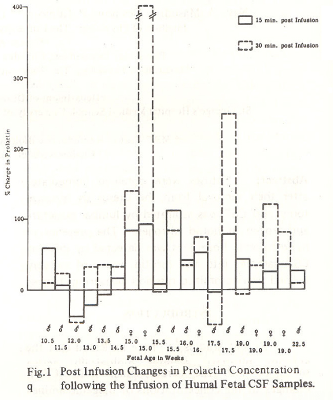
Statistical analysis of these observations showed significant prolactin releasing activity in the CSF samples. The infusions of male and female fetal CSF samples both caused a significant increase in serum prolactin concentrations.
The presence of LHRH activity was also detected in human fetal CSF samples of mid-term gestational age. Twenty - CSF samples were gestational age. Twenty CSF samples were examined using the same experimental procedure as followed for prolactin releasing activity determinations. LHRH activity was demonstrated by the rise in circulating LH brought about by the infusion of CSF. The results obtained in this study (Fig. 2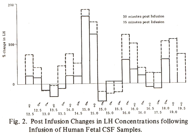
clearly confirmed the presence of LHRH activity in human fetal CSF collected from both male and female fetuses. Significantly higher (P < 005) serum LH concentrations were obtained in the post infusion 15 minutes blood samples in both sexes. This rise became even30 minutes after more significant (P < 0.00 1) the infusions were carried out.
DISCUSSION
It is now widely accepted amongst neuroendocrinologists that brain-pituitary relationships in the human are more complex than had previously been considered. Since hypothalamic hormones and biogenic arnines can be detected in the CSF, it is natural to speculate that the CSF has an important role to play in carrying information between the brain and pituitary9. Very little information is, however, available regarding the presence of hypothalamic hormones and biogenic amines in CSF collected from mid-term human fetuses.
The possible role of the CSF in carrying hypothalamic releasing and/or inhibitory hormones towards the pituitary via the third ventricle has been reported by a number of workers13,14 A demonstration in this study of the presence of LHRH and prolactin releasing activity in human fetal CSF confirms some of the previous work carried out in the adult human CSF12,19-24
Intraspinal TRH injections in the human produce a significant rise in blood thyrotrophins and prolactin levels25. Thus it could be assumed that the natural release of TRH and LHRH into the human fetal CSF may be having a similar effect. These hormones could diffuse into the pituitary and so affect particular pituitary cells to produce their respective trophic hormone. This view is further supported by another similar observation in rats. LHRH injected into CSF was found to significantly elevate plasma LH Levels16,26.
It is well known that until some time between the 18th and 20th week of gestation, the hypothalamic anterior pituitary portal connections are not well established in the fetus27,28 Therefore, the CSF may, prior to this time, have a major role in transporting LHRH from the hypothalamus to the anterior pituitary. The presence of LH in the fetal pituitary gland as early as 10 weeks gestational age and equally-early the presence of LHRH in the hypothalamus29 imply the potential for an early hypothalamic pituitary functional relationship. The presence and activity of LHRH seen in fetal CSF in this study suggests the CSF does have a role in this functional relationship.
The results of the present study also clearly indicate that prolactin releasing activity (like that of LHRH) may be effected by either:
(a) a direct action of the hypothalamic hormones at the level of the pars tuberalis and the tuber cinereum, which are distinctly vascularized from an early stage30, or
(b) indirectly through transport in the CSF to the infundibular recess into the pars distalis.
ACKNOWLEDGEMENTS
We thank Professor M.C. MacNaughton, Dr. Man Calder and their staff at Glasgow Royal Infirmary for enabling us to obtain the hysterotomy specimens; Miss Alison McCulloch, Miss Georgina Docherty for their excellent technical assistance and Mrs. Moira McRae for typing this manuscript.
This work was supported by grants from the British Council, London under Technical Cooperation Training Development Programme under Colombo Plan.
REFERENCES
1. Bergland, R.M., Davis, S.L. and Page, R.B. Pituitary secretes to brain; experiments in sheep. Lancet, 1977;2 :276.
2. Bergland, R.M. and Page, R.B. Pituitary-brain vascular relations; a new paradigm. Science, 1979; 204 : 18.
3. Rodriguez, EM. The cerebrospinal fluid as a pathway in neuroendocrine integration. J. Endocrinol., 1976;71 :407.
4. Kendall, J.W., Seaich, J.L., Allen, J.P. and Vanderlaa, W.P. Pituitary-CSF relationships in man. In K.M. Knigge, D.E. Scott and A. Weindle (Eds), Brain-endocrine Interaction. II. The Ventricular System, Karger, Basel (1975). 313-323.
5. Knigge, K.M., Joseph, S.A.. Sladek, J.R., Notter, ME., Morris, M., Sundberg, D.K., Holzwarth, M.A., Hoffman, G.E. and O’Brien, L. Uptake and transport activity of the median eminence of the hypothalamus. Int. Rev. Cytol., 1976;45 :283.
6. Mezey, E., Palkovits, M., de Kloet, E.R., Verhoef, J. and deWeid, D. Evidence for pituitary brain transport of a behaviorally potent ACTH analog. LifeSci., 1978;22 :831.
7. Oliver, C., Mical, R.S. and Porter, J.C. Hypothalamic pituitary vasculature; evidence for retrograde blood flow in the pituitary stalk. Endocrinology, 1977;101 :598.
8. Assies, J., Schellekens, A.P.M. and Touber, J.L. Prolactin in human cerebrospinal fluid. J. Clin. Endocrinol.Metab., 1978;46 :576.
9. Hyyppa, M.T. and Liira, J. Neuropeptide hormones in cerebrospinal fluid; experimented and clinical aspects. Med. Biol., 1979;57 :367.
10. Miyake, A., Kurachi, H., Kawamura, Y., Aono T.,Kurachl, K. and Yanagida, T. Luteinizing hormone-releasing hormone in cerebrospinal fluid of women. Endocrinol. Jpn., 1980;1: 117.
11. Oliver, C., Ben-Jonathan, N., Mical, R.S. and Porter, J.C. Transport of thyrotropin-releasing hormone from cerebrospinal fluid to hypophysial portal blood and the release of thyrotropin. Endocrinology, 1975;97 : 1138.
12. Young, S.N., Anderson, G.M., Gauthier, S. and Purdy, W.C. The origin of indoleacetic acid and indolepropionic acid in rat and human cerebrospinal fluid. J. Neurochem., 1980; 34:1087.
13. Young, S.N., Anderson, G.M. and Purdy, W.C. Indoleamine metabolism in rat brain studied through measurements of tryptophan, 5 -hydroxyindoleacetic acid, and indoleacetic acid in cerebrospinal fluid. J. Neurochem., 1980; 34 : 309.
14. Findlay, A.L.R. Hypothalamic inputs; methods and five examples. Prog. Brain Res., 1972; 38:163.
15. Hayward, J.N. Hypothalamic input to supraoptic neurones. Prog. Brain Res., 1972; 38 145.
16. Porter, J.C., Ben-Jonathan, N., Oliver, C. and Eskay, R.L. Secretion of releasing hormones and their transport from CSF to hypophysial portal blood. In K.M. Knigge, D.E. Scott and A. Weindi (Ms.) Brain-endocrine Interaction. II. The Ventricular System, Karger, Basel (1975) pp. 295 -305.
17. Iffy, L., Jakobovits, A., Westlake, W., Wingate, M., Caterin, H., Kanafsky, P. and Menduke, M. Early intrauterine development I. the rate of growth of Caucasian embryos and fetuses between the 6th and 20th weeks of gestation. Pediatrics, 1975;56 : 173.
18. Gilmore, D.P., Dobbie, H.G., McNeiily, A.S. and Mortimer, C.H. Presence and activity of LH-RH in the mid-term human fetus. J. Reprod. Fert., 1978;52 : 355.
19. Knigge, K.M. and Joseph, S.A. Thyrotrophin releasing factor (TRF) in cerebrospinal fluid of 3rd ventricle of rat. Acta. Endocrinol. (Copenh.), 1974; 76 : 209.
20. Liira, J., Leppaluotto, J. and Hyyppa, M.T. Hypothalamic and pituitary hormones in human CSF. Acta. Neurol. Scand., 1978; 57 (SuppL 67): 272.
21. Loose, R. and Paterson, W.G. 5-hydroxyindole acetic acid in amniotic fluid and foetal 5-hydroxy-tryptamine metabolism. J.Obstet. Gynaecol. Br. Common., 1966;73 :647.
22. Oliver, C., Charvet, J.L., Codaccioni, J .L., Vague, J.and Porter, J.C. TRH in human CSF. Lancet, 1974;1 :873.
23. Patel, Y.C., Rao, K. and Reichlin, S. Somatostatin in human cerebrospinal fluid. N. Engl. J. Med., 1976;296 :529.
24. Vaala, S.S. and Knigge, K.M. Transport capacity of median eminence; in vitro uptake of 3H-LRF. Neuroendocr., 1974; 15 : 147.
25. Vigneri, R., Pezzino, V., Filetti, S., Squatrito, S., Corso, A., Maricchiolo, M., Polosa, P. and Scapa gnini, U. Thyrotropin and prolactin response to intraspinal TRH administration in man. Neuroen docrinology, 1977; 23 :171.
26. Ondo, J.G., Esay, R.L., Mical, R.S. and Porter, IC. Release of LH by LRF injected into the CSF; a transport role for the median eminence. Endocrinology, 1973;93 :231.
27. Raffia, N. and Hjelt, L. The correlation between the development of the hypophysial portal system and onset of neurosecretory activity in the human fetus and infants. Acta Paediat. Upps., 1957; 46 :610.
28. Rinne, U.K. Neurosecretory material passing into the hypophysial portal system in the human in fundibulum, and its foetal development. Acta Neuroveg. (Wien), 1963;25 :310.
29. Kaplan, S.L., Grumbach, M.M. and Aubert, M.L. The ontogenesis of pituitary hormones and hypothalamic factors in the human fetus; maturation of central nervous system regulation of anterior pituitary function. Recent Prog. Horm. Res., 1976;32 :161.
30. Atwell, W.J. The development of the hypophysis cerebri in man, with special reference to the pars tuberalis. Am. J. Anat., 1926; 37 : 159.
Journal of the Pakistan Medical Association has agreed to receive and publish manuscripts in accordance with the principles of the following committees:


