

Shafqat Ullah Memon ( Department of Medicine, Jinnah Postgraduate Medical Centre, Karachi. )
S.M. Rab ( Department of Medicine, Jinnah Postgraduate Medical Centre, Karachi. )
September 1991, Volume 41, Issue 9
Case Reports

Neuro-osteoarthropathy (Charcot’s Joint) in diabetic patients has been well described in literaturc1-9. It has been defined as a chronic progressive degenerative process principally occurring in weight-bearing joints in the feet of diabetic patients who have underlying peripheral neuropathy and loss of sensation6,9,10. Although considered as a clinical rarity3,6, it has been reported more frequently in recent years7-9,11,127-9,11,12. This observation can be attributed to the longevity of diabetic patients and dramatic decrease in mortality since the insulin era7.
The association between juvenile onset diabetes and skeletal lesions is relatively uncommon12,13. We report a case of young woman who had juvenile onset diabetes and developed bilateral neuro-arthropathy involving feet.
Case Report
A 23 year old female, known diabetic for 8 years and on insulin therapy was admitted in January, 1990 with uncontrolled diabetes and severe hypertension. She complained of swelling of both feet for the past 2 years but denied having sustained any trauma to them. Clinical examination revealed a blood pressure of 210/130 mm Hg and fundal changes consistent with diabetes and hypertension. Both feet appeared swollen with slight tenderness. Neurological examination revealed decreased sensation to pinprick and bilaterally impaired sensation of position and vibration. Her ankle and knee jerks were absent on both sides. Pulses were palpable in both femoral and popliteal arteries. Dorsalis pedis and posttibial appeared normal. Investigations revealed an elevated blood urea (114 mg %) and creatininc (2.6 mg %). Radiographs of feet showed slight flattening of calcaneus and a crack in the bony bridge between calcaneus and navicular bone on the left side, while on
the right side there was beginning of disorganization of the subtalar joint with loss of depth of the body of talus. (Figures 1,2).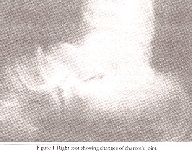
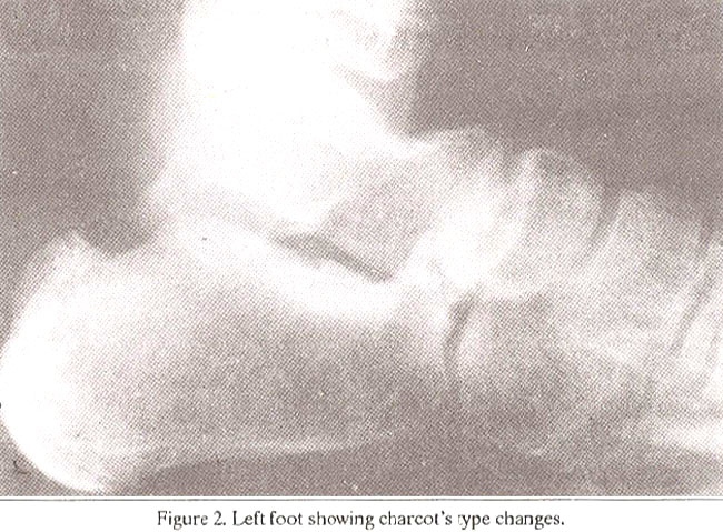
Her diabetes was controlled on insulin and blood pressure reduced and maintained on medication. For her feet she was instructed complete bed rest, non-weight bearing and special protective shoes. She was discharged after control of her blood pressure and was advised periodic follow-up but she did not report back and died 3 months later of renal failure.
DISCUSSION
Since Charcot’s initial report of neuropathic joints in association with tabes1,4,8,10,12,14. a multitude of underlying causes have been ascribed to the development of this lesion, the predominant being syringomvelia, leprosy, spina bifida, cord trauma, multiple sclerosis and spinal dysraphism1,2,4,7-9,15-16 Diabetes mellitus, however, remains the commonest causeS, 16 and its associated bone and joint abnormalities are nowwell recognized2,3,5,7,10,12,13
The reported incidence varies between 0. 1 to 6.8%5,8,18-20. Majority of the cases are of adult onset diabetes often non- insulin dependent13,14,16 lesions occurring over the age of fifty5,6,13,14,16 after the Patient has had diabetes for an average of 1 3to 15 years 6,13 The blood sugar levels are often poorly controlled 5,68 although osteopathy may even precede overt diabetes5. While the high risk group constitutes those with characteristic triopathy6,12, the diabetics with renal transplants are at greatest risk12,21. More recently an association between joint lesions and pulmonary changes in diabetics has been described22-24. Therefore, patients with impaired respiratory funct ion can constitute another high risk group.
Typical neuropathic joints in diabetics can be seen in any pari16,25-27 hut the foot remains the area most frequently involved14 The sites most often affected are the tarsometatarsal joints (60%) and to a lesser extent the joints of metatarso-phalangeal area (30%) and ankle (10%)6,9,18. A clear distinction has been made between proximal foot destruction and distal foot erosion7,10. On roentgenogram two forms exist - ATROPIIIC and HYPERTROPHIC, the latter being described as osteophytic fragmentation,joint space narrowing, capsular distention and lipping that predominantly affects the lower extremities. The atrophic form commonly affects the upper extremities and the only change is resorption of bone ends1,2,6,9,16.. Transition from one form to the other does not occur6,16. Extensive periartieular calcification is characteristic28,29 occasionally bone shards may be seen far from the joint suggesting their denovo origin from the joint capsule29.
The pathophysiology of Charcot’s joint remains controversial, In attempts to elucidate pathogenesis various mechanisms were described pertaining to trophic disturbances, trauma1,5,6,8 autonomic impairment30, infection31, isehaemia32, bone atroph33, OS teoporosis5,6, small vessel disease34, and to an error of protein metabolism3. However no explanation was based on conclusive evidence that focussed on any single process. Current concepts suggest that osteolysis and bone resorption play a major role in the development of Charcot’s joints33-36. and for this an adequate blood supply is a prerequisite37,38. The clinical finding of bounding pulses39-41 and the development of Charcot’s joint in ischaemic limbs following revascularization surgery9 strongly support this hypothesis. Edelman et al9 have suggested that while peripheral neuropathy is the initial primary defect, a neurally initiated vascular reflex leading to increased blood flow may play a pivotal role in the development of Chareot’s joint. Mechanical trauma in an insensitive foot is contrihutary but a secondary mechanism9,
Whatever the mechanism, a marked disproportion between the radiologic severity of the disease process and the relatively slight complaint of discomfort is thc most consistent clinical feature. Often there is no history of trauma and, when inflicted, may be trivial. This explains why the lesions remain unreeognised for long and the changcs are far advanced when first presented18.
Our patient clinically had a classical Charcot’s joint. She had severe clinical manifestations of diabetes and represented the high risk group The lesion developed without known or obvious trauma and at the time of presentation her blood sugar levels were poorly controlled. Nevertheless the case is unusual in several aspects. The patient was rather young and had juvenile onset insulin dependent diabetes which is relatively uncommon in patients developing neuropathic joints. Moreover, the lesion appeared rather early. Harris & Brand10, observed five patterns of disintegration in the anaesthetic foot and our case resembled type TI thcy described as central destruction which is infrequently encountered, as it is not common for talus to be the primary focus of disintegration. The pathogenctic factors in our ease remain speculative. Where- as the destruction of talus seems to he entirely due to mechanical stress10, the presence of unimpaired peripheral circulation favours the hypothesis of osteolysis and bone resorption9. Probably both these factors in a neuropathie limb favour the development of Chareot’s joint.
Diabetic neuro- osteoarthropathy has a characteristic intermittent course of activity5. Trauma unquesuonably causes deterioration5,6. Current treatment protocol,therefore, includes non- weight-bearing and immobilization of the involved extremity and prophylactic immobilization of contralateral extremity. Control of blood sugar levels is desirable and serial, roentgenograms are advised to monitor progress6,8,9,11,12. In absence of marked bony deformities the long term prognosis is generally favourable6.
REFERENCES
1. Delano, PJ. The Pathogenesisof Charcot’s joint. AJR., 1916:56:189 189.
2. Gould, D.M. and McAfee, J.G Roentgenographic manifestations of diabetes mellitus. Am.). Med. Sci., 1960; 239:622.
3. Pogonowska, M.J., Collins, L.C. and Dobson, HI... Diabetic osteopathy. Radiology. 1967; 89:265.
4. Johnson. J.T. Neuropathie fraeturesand joint injuries.). BoneJointSurg., 1967; 49-A:1.
5. Gondos, B. Roentgen observation in diabetic osteopathy. Radiology, 1968; 91:6.
6. Sinha, S., Munichoodappa, C.S. and Kozak, G.P. Neuro-arthropatlsy (Charcotjoints) in diabetes meltitua. Clinical study of 101 cases. Medicine, 1972; 51:191.
7. Kraft, E., Spyropoulos, B. and Finhy. N. Neurogenie disorders of the foot in diabetes mellitua. AIR., 1975; 124:17.
8. Essea, S., Langer, F. and Gross, A. Cbarcot’s joint. A ease report in a youngpatientwith diabetes. Clin. Orthop., 1981; 156:183.
9. Edelman, S.V., Koaofsky, EM., Paul, R.A. and Kozak, G.P. Neuro- osteoarthropathy (Charcot’s joint) in diabetes mellitus following revascularization surgery. Three cases report and a review of the literature. Arch. Intern. Med., 1987; 147:1504.
10. Harris, J.R. and Brand, P.W. Patterns of disintegration of the tarsus in the anaesthetic foot). Bone JointSurg., 1966; 48- B:4.
11. El-Khoruy, G.Y. and Kathol, M.H. Neuropathic fractures in patients with diabetes mellitua. Radiology, 1980; 134:311
12. Clohisy, D.R. and Thompson, R.C. Fractures associated with neuropathie arthropathy in adultswho havejuvenile onset diabetes.). Bone Joint Surg., 1988; 70-A:1192.
13. Forgaca, S. Clinical picture ofdiabetic osteoarrhropathy. Acts Diabet Lat, 1976; 13:111, t4. Newman, J.H. Spontaneous dislocation in diabetic neuropathy. A report of six cases.). Bone Joint Surg., 1979; 61-8:484.
15. Nellhaus, G. Neurogenic arthropatlsies (Charcot’s joints) in children. Description of a case traced to occultspinal dysrophiam. Clin. Pediatr., 1975; 14:647.
16. Schwarz, G.S., Berenyi, M.R. and Siegel, MW. Atrophic arthropsthy and diabetic neuritis. MR., 1969; 106:523.
17. Coventry, MB. and Rothscker, G.W. Bilateral eslcaneal fracture ins diabetic patient). Bone Joint Surg,, 1979; 61-A:462.
18. Frykberg, R.G. and Kozak, UP. Neuropathic arthropathy in the diabetic foot Am. Fsm. Physician, 1978; 17:105.
19. Bailey, C.C. and Root, H.F. Neuropsthic foot lesions in diabetes mellitus. N. Engi. 3. Med., 1947; 236:397.
20. Martin, M.M. Neuropsthic lesions of the feet in diabetes mellitus. Proc. R. Soc. Med., 1954; 47:139.
21. Thompson. R.C. Jr., Hsvel, P. and Goetz, F. Presumed neurotrophicskeletal disease in diabetic kidney transplant recipients. JAMA., 1983; 249:1317.
22. Buckingham, B., Perejda, A.)., Sandborg, C. et at Skin, joint & pulmonary changes in type I diabetes mellitus. Am.). Dis. Child., 1986; 140.420.
23. Msdscsy, I..., Peja, M., Korompay, K, et al. Limited joint mobility, impaired respiratory function & early retinopsthyin childhood diabetes. Diabetologia 1982; 23:184.
24. Madscsy, L, Pejs, M., Kornmpsy, K. and Biro, B. Limited joint mobility in diabetic children; a risk fsctorof diabeticcomplications? Acts Pscdistr. Hung., 1986; 27:91.
25. Zucker, U. and Marder, M.J. Chsrcot spine due to diabetic neuropsthy. Am. 3. Med., 1952; 12:118
26, Feldman, S., Johnson, AM. and Walter, J,F. Acute sxisl neurosrthropsthy. Rsdiology, 1974; 111:1.
27. Feldman, M.J, Becker, K.L., Reefe, W.E. snd Longo, A. Multiple neuroparhic joints, including thewrist, in a pstientwith diabetes mellitus. JAMA., 1969; 209:1690.
28. Hotwitz, T. Bone and cartilage debris in the synovial membrane. J.Bone Joint Surg,, 1948; 30-A:579.
29. Forrester, D.M.and Msgre. U. Migratingbone shards in dissectingCbarcotjoint. MR., 1978; 130:1133.
30. Foster, D.B. and Bassett, R.C. Neurogenic arthropathy (Chsrcot joint) associated with diabetic neuropathy. Arch. Neurol. Psychiatry, 1947; 57:173.
31. Hodgeon, J.R., Pugh, D.G. and Young, H.H. Roentgenologic aspects of certain lesions, of bone. Neurotropic or infectious? Radiology, 1948; 50:65.
32. Psrsons, H. and Norton, W.S. Management of diabetic neuropathic joints. N. EngI. J, Med., 1951; 244:935.
31 Gillespie, IA. The nature of the bone changes associated with nerve injuries and disuse.
32. Bone Joint Surg., 1954; 36-B:464.
34. Pedersen, 3. and Olsen, S. Small.veasel disease of the lowerextremity in diabetes mellitus. On the psthogeneaia of the foot’lesions in diabetics. Acts Med. Scand., 1962; 171:551.
35. Delbridge, L., Ctercteko, U., Fowler, C., Reeve, T.S. and LeOuesne, LP. The etiotogyof disbetic neuropsthic ulceration of the foot Br.J. Surg., 1985; 72:1,
36. Brooks,A,P. Theneuropsthic footin diabetes. II. Charcot’s neuroarthropsthy. Diabetic Med., 1986; 3:116.
37. Phemister, D.B. Lesions of hones and joints arising from interruption of circulation.). Mt Sinai Hosp., 1948; 15:55.
38. Scott,). Bone changes in chronic arterisl insufficiency. Angiology, 1959; 10:382.
39. Ftykberg, R.G and C.P. The diabetic Charcot foot, in management of Diabetic Foot Problems. Edited by Kozak UP, Hoar CS, Philadelphia, Saunders, 1984, p. 103.
40. Goldman, F. Indentification, treatment, and prognosis of Charcot joint in diabetes mellitus. 3. Am. PodistryAssoc., 1982; 72:485.
41. Frykberg, R.G. Neuropsthic arthropathy; the diabetic Charcot foot Diabetes Educ., 1984; 9:17.
42. Gristins, A.G.,Nicsstro, 3., Clippinger, F. and Rovere, GD. Neuropathy of the foot and ankle. J.Bone Joint Surg., 1975; 57-A:1035.
Related Articles
Journal of the Pakistan Medical Association has agreed to receive and publish manuscripts in accordance with the principles of the following committees:


