
G.M. Memon ( Department of Pathology, Basic Medical Sciences Institute, Jinnah Postgraduate Medical Centre, Karachi. )
S.M.Alam ( Department of Pathology, Basic Medical Sciences Institute, Jinnah Postgraduate Medical Centre, Karachi. )
February 1993, Volume 43, Issue 2
Original Article
ABSTRACT
Monoclonal antibody Leu M1 represents a highly specific marker to locate granulocyte antigen in Reed-Sternberg cells in Hodgkin’s disease. Except the L&H variants of reed-sternberg cells in lymphocyte predominance variety in which antigens are probably sialylated. All the cases of non-Hodgkin’s lyniphoma were negative with this marker, because of absence of antigen. Therefore this specific marker characterizes granulocyte origin of reed- sternberg cells (JPMA 43: 32, 1993).
INTRODUCTION
Malignant lymphomas are recognized as clonal proliferations of specific elements of the immune system. The cellular origin of Hodgkin’s disease has, however, remained an enigma. The reed- sternberg cells and related mononuclear cells are established as the neoplastic elements of Hodgkin’s disease, but the derivation of these cells remains speculative. Evidence has suggested origins from monocyte/macrophage1-3. T and B lymphocytes4-6, dendritic reticulum cells7,8, granulocytes9 and uncharacterized lymphnode mononuclear cells10. Recent studies with monoclonal antibodies have demonstrated antigenic similarities to granulocytes11. In the present study we examined the antigenic expression of reed-sternberg cells in 50 cases of Hodgkin’s disease, utilizing Leu M1 (antigranulocyte) monoclonal antibody. We also examined 31 cases of non-Hodgkin’s lymphoma to see the antigenic expression of neoplastic cells. The results obtained are being reported here.
MATERIALS AND METHODS
Tissues:
Eighty-one specimens of lymphnode biopsies received in the department of pathology, during the period 1st January, 1983 to 31st October, 1988, diagnosed on H&E staining were selected for study. Fifty cases were of Hodgkin’s disease and 31 ofnon- Hodgkin’s lymphoma. Monoclonal antibody Leu M1 (Becton and Dickinson), universal immunoperoxidase kit (Dakopatts), trypsin (Sigma Corporation) and AEC (3-amino-9 ethyl carbazole) was used as chromogenic substrate. Tissue sections were mounted on poly-L-lysine coated slides, deparaffinized through xylene and rehydrated through graded alcohol and placed in phosphate buffered saline (PBS) followed by methanolic hydrogen peroxide for 30 minutes at room temperature to reduce non-specific background staining by blocking endogenous peroxidase. The slides were then washed and placed in PBS. The sections were incubated in a solution of trypsin for 20 minutes at 37’C for proteolytic digestion. The slides were washed and placed in PBS. The sections were treated with normal swine serum for 45 minutes at 37°C to avoid non-specific staining. The sections were treated with primary antibody (mouse antthuman) Leu M1 (1:50) and incubated in humidity chamber for 1.15 hour at 37°C. The slides were washed in three changes of PBS for 15 minutes. The sections were treated with secondary antibody (peroxidase conjugated rabbit antimouse immunoglobulin) and incubated in humidity chamber for 45 minutes at 37°C. The slides were washed in three changes of PBS for 15 minutes. The sections were treated with tertiary antibody (peroxidase conjugate swine antirabbit immunoglobulin) and incubated in humidity chamber for 45 minutes at 37°C. The slides were washed in three changes of PBS for 15 minutes. The sections were treated with AEC chromogenic substrate solution and incubated for 40 minutes in humidity chamber at room temperature. The slides were rinsed with distilled water and then counterstained with Mayer’s hematoxylin for 5 minutes. The slides were rinsed with distilled water. The sections were flooded with ammonia water and incubated for 10 seconds to develop counterstain. The excess water was wiped off and coverslips were mounted by using glycerine jelly. The positive control slides were prepared from tissue of acute suppurative appendicitis containing polymorpho nuclear leukocytes and stained in similar manner. The negative control sections were stained with omission of primary antibody and replaced by normal non-immune serum.
RESULTS
All the cases of Hodgkin’s disease, which were included in the study showed positive staining with Leu M1 (antigranulocyte) monoclonal antibody except lymphocyte predominance variety, suggesting the presence of granulocyte related antigens in the reed-sternberg cells (Table). None of the cases of non-Hodgkin’s lymphoma showed positive staining with antigranulocyte monoclonal antibody, suggesting the absence of granulocyte related antigens in the neoplastic cells (Table).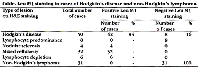
DISCUSSION
All cases of Hodgkin’s disease except lymphocyte predominance variety showed positive and cases of non-Hodgkin’s lymphoma showed negative staining with Leu M1 antibody because atypical histiocytes of these cases did not have Leu M1 antigen. A number of studies showed that except for lymphocyte predominance variety, all the cases of Hodgkin’s disease were positive with Leu M1 marker12-17. The lymphocyte predominance variety were positive with Leu M1 marker when these cases were pretreated with neuraminidase enzyme which removes sialic acid from L&H variants of reed-sternberg cells14. Furthermore antibodies that react relatively with antigen on the cells involved in the later stages of normal granulopoiesis (antigranulocyte antibodies) also recognized the tumour cells in most cases of Hodgkin’s disease. This may shed new light on the origin of H&SR cells. The presence of granulocyte cell specific antigens in H&SR cell is not consistent with the concept of histiocyte or lymphoid origin9. Several investigations have shown that reed-sternberg cells contain both kapa and lambda chains whereas normal and neoplastic B cells contain either kapa or lambda but not both. Furthermore with the exception of some cases of lymphocyte predominance variety H&SR cells constantly lackJ chain which are frequently observed in B-cell malignancies9. The finding of trace production of lysozyme by cultured H&SR cells and the presence of alpha-1-antitrypsin would also favour granulocytic origin because of many granulocytic cells contain lysozyme and alpha-1-antitryp-sin18,19. Studies of Hodgkin’s disease with panel of monoclonal antibodies also favoured its granulocytic derivation20. Leu M1 marker is a specific one to locate granulocytic antigens in H&SR cells in Hodgkin’s disease. This marker did not stain giant cells resembling R-S cells in non-Hodgkin\\\'s lymphoma because of the absence of granulocytic antigen. Leu M1 did not stain L&H variants of R-S cells in lymphocyte predominance variety of Hodgkin’s disease because granulocyte antigen in these cells is probably sialylated. Our results conform to others findings that Leu M1 marker suggests the granulocytic origin of H&SR cells.
REFERENCES
1. Kadin, M.E., Stites, D.P., Levy, R. and Warner, R. Exogenous immunoglobulin and the macrophage origin of reed-sternbergcells in Hodgkin’s disease. N. EngI. J. Med., 1978; 299:1208-14.
2. Kaplan, H.S. and Gartner, S. Reed-aternberggiant cellain Hodgkin’s disease; cultivation in vitro, hetrotransplantation and characterization of neoplastic macrophages. Int. J. Cancer, 1979; 19:511-25.
3. Strauchen, J.A. Lectin receptors as markers of lymphoma cells II. Reed-sternberg cells share lectin-binding properties of monocyte macropltiges. Am. ). Pathol., 1484; 116:370-76.
4, Gravin, A.)., Spicer, 5.5., Parmley, R.T. and Munater, AM. lmmunohiatochemical demonstration of IgG in reed-sternberg and other cells of Hodgkin’s disease. J. Exp. Med., 1974;134:1077-83.
5. Glick, AD., Leech, ).H., Flexner, J.N. and Collina, RD. Ultraatructural study of reed-sternberg cells. Am.). Pathol., 1976;85:195-208.
6. Biniaminov, M. and Ramot, B. Possible T-lymphocyte origin of reed-sternberg cells. Lancet, 1974;1:368.
7. Kadin, M.E. Possible origin of the reed-sternberg cells from an interdigitatingreticulurn cell. CancerTreat. Rep., 1982;66:601-8.
8. Becatead, ).H., Warnke, R. and Bainton, D.F. Hiatochemiatry of Hodgkin’s disease. CancerTreat. Rep., 1982;66:601-8.
9. Stein, H., Uchanaka-Ziegler, a, Gerdea, )., Ziegler, A and Wernet, P. Hodgkin and sternberg-reed cells contain antigens specific to late cells of granulopoeiaia. Int. i. Cancer, 198Z29:283-90.
10. Stein, H., Gerdes, 3., Schwab, U., Lemke, H., Mason, D.Y., Ziegler, A, Schienle, W. and Diehl, V. Identification of Hodgkin and sternberg-reed cella as a unique cell type derived from a newly-detected small-cell population. Int.J. Cancer, 1982;30:445.59.
11. Norton, A). and Isaacaon, P.O. Granulocyte and HL.A-D region specific monoclonal antibodies in the diagnosis of Hodgkin\\\'s disease. J.Clin Pathol., 1985;38:1241-46
12. Hsu, S. and )affe, ES. Leu M1 and peanut agglutinin atain the neoplaatic cells of Hodgkin’s disease. Am.). Clin. Patbol., 1984;82:29-32.
13. Hau, M., Yesten, H., Khalil, Sand Winberg, C.D. L&H varianta of reed sternberg cells express sialylated Leu ml antigen. Am.).Clin. Pathol., 1986;122:99.
14. Pinkus, OS. and Said, J.W. Hodgkin’s disease. Lymphocyte predominance type, nodular-A diatinctentity? Am.). Pathol., 1985;118:1-6.
15. Strauchen, ).A. and Alexandria Dimitriubona, A. Immunopathology of Hodgkin’s diseaae. Characterization of reed aternberg cells with monoclonal antibodies. Am.). Pathol., 1986;123:293-300.
16. Hsu, S., Yang, K. and Jaffe, E.S. Phenotypic expression of Hodgkin\\\'s and reed-sternberg cells in Hodgkin’s diseaae. Am.). Pathol, 1985;118:209-17.
17. Swerdlow, S.H. and Wright, S.A. The apectrum of Leu ml staining in lymphoid and hematopoietic proliferationa. Am.). Clin. Pathok, 1986;85:283-88.
18. lsaacson, P. and Wright, D.H. Anomalouastaining patternain immunohiatologicatudies of malignant lymphoma. 3. Hiatochem. Cytochem., 1979;27:1197-99.
19. Poppema, S.The diversityof the immunohiatological staining pattern of sternberg-reed cells.). Hiatochem. Cytochem., 1980;28:788-91.
20. Zeigler, A, Diehl, V., Uchanaka-Zeigler, B., Kirchner, H., Burrichter, H.and Wernet, P. Cell aurfaceantigena on a Hodgkin’s disease derived cell line, L 428 cells and 5 aubclones aa detected by monoclonal antibodies. Immunobiology, 1981;160:137-38.
Journal of the Pakistan Medical Association has agreed to receive and publish manuscripts in accordance with the principles of the following committees: