

Muhammad Muzaffar ( Department of Histopathology, Armed Forces Institute of Pathology, Rawalpindi. )
Nadira Mamoon ( Department of Histopathology, Armed Forces Institute of Pathology, Rawalpindi. )
Ezra Nigar ( Department of Histopathology, Armed Forces Institute of Pathology, Rawalpindi. )
Sajid Mushtaq ( Department of Histopathology, Armed Forces Institute of Pathology, Rawalpindi. )
August 1997, Volume 47, Issue 8
Case Reports

Introduction
Endometrial ossification is a rare condition. The presence of bone in the uterus is explained on the basis of several debatable hypotheses1-3. We report three such cases with different clinical presentations suggesting varied pathogenesis for this uncommon finding.
Case Report
Case 1
A 38 years old multiparous lady presented with an eight month history ofpolymenorrhagia. The patient gave history of abortion at 8 weeks gestation, an year back. Despite conventional hormone therapy the patient’s complaints persisted. A hysterectomy with right sided salpingoophorectomy was performed. Gmss examination of the specimen showedanormal sized uterus. The uterine cavity was found to be full of tiny fragments of bone adherent to and lining the endometrial cavity. The cervix was found to be normal. The ovary and tube were also unremarkable. Histological examination showed fragments of maturebone in the endometrium. The glands were in the proliferative phase and lymphocytic infiltration of the stroma and glands was seen. The cervix, ovary and tube were nonnal.
Case 2
A 20 year old lady presented with unexplained primary infertility of 5 years duration. No abnormality was detected on physical or pelvic examination. A diagnostic curettage was perfonned. Histology showed late secretory endometrium with fragments of mature bone in the stroma. No evidence of chronic endometritis was seen (Figure).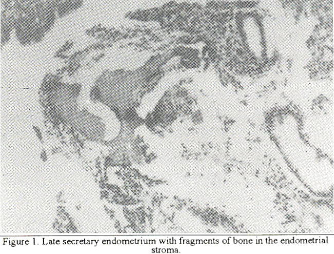
Case 3
A 22-year old lady presented with a history of primary infertility for the last five years. No abnormality was detected on physical examination. Ultrasound showed the presence of bone inthe uterine cavity. A diagnostic curettage was done. On histology, a proliferative phase endometrium was seen alongwith extensive calcification and ossification of the stroma. The stroma and glands were also densely infiltrated by polymorphs, lymphocytes and plasma cells. A diagnosis of non-specific endometritis with osseous metaplasia of endometrium was made in this case.
Discussion
Endometrial ossification is an uncommon finding and only a limited number of cases have been documented in the literature1, Several suggestions regarding the pathogenesis of this lesion have been made. Chronic endometritis accompanied by osseous metaplasia is considered to be one mechanism2,4,5. Dystrophic calcification and ossification of retained fetal tissue after second - trimester abortion has also been described6. Another hypothesis is that heteroplasia occurs in multipotential stromal cells, probably of Mullerian origin, that lie silently in the uterus in the myo-endometrial zone3,6,7. The bony fragments in our first case were visible both macroscopically as well as microscopically. A past history of abortion followed by polymenorrhagia is strongly suggestive of ossification of retained fetal tissue. Since the termination of pregnancy occurred at 8 weeks gestation, the bony fragments cannot be of fetal origin, as suggested by some observers6. These bony spicules were found only in the superficial layers of the endometrium and had a well differentiated structure. The bony fragments act as a foreign body in the uterus and may lead to menorrhagia, pelvic pain and vaginal discharge. We believe that our second case presenting with primary infertility is due to osseous mctaplasia of multipotcntial stem cells, since no evidence of chronic endometritis was found in the curettings. The bony fragments may lead to infertility either by altering the milieu of the uterine cavity and thereby increasing prostaglandin production or if large and superficial enough, by acting as an intra-uterinc contraceptive device and prevent implantation3. Our third case showed severe chronic endometritis with osseous metaplasia. Here the bone comprised anastomosing trabeculae of well fonned bone alongwith extensive calcification. Whether this was secondary to chronic endometritis or whether chronic infection was itself a secondary event to the presence of bone in the uterus could not be ascertained. While investigating patients of post-abortion menorrhagia or primary infertility, the possibility of endometrial ossification should always be kept in mind. The role of pelvic ultrasonography, in this regard, cannot be over-stressed. Hysteroscopic removal of bony fragments may relieve symptoms in menorrhagia and may restore fertility3.
References
1. Marus, S.F, Bhattarcharya, J.. Williams, 0. eta!. Endometrial ossification: A cause of secondary infertility - Report of two cases. Am. J. Obstet. Gynecol. 1994;170:1381-1383.
2. Camus, M., Report, J.F, Iloki. L.H. eta!. Endometrial ossifleation. Apropos of 5 recent cases. J. Gynecol. Obstet. Biol. Reprod. (Paris), 1990;19:295-300.
3. Bhatia, N.N. and Hoshiko, MG. Uterineosseous metaplasia. Obstet. Gynecol., 1982;60 :256-9.
4. Ejeckam, G.C., Haseeb, F., Ahmad, R. et sl. Endometrial ossification. Trop. Geogr. Med., 1991;43:314-316.
5. Rodrigvez, B.D. snd Adamson, GD. Hysteroscopic treatment of ectopic intrauterine bone, Acasereport. 3. Reprod. Med., 1993;38:515-20.
6. Hoang, N.M., Lemay, B. and Smsdja, A. Reflections on endometrial osteogenesis. Apropos of 3 eases. Rev. Fr. Gynecol. Obstet., 1984;79:471-474.
7. Verstrsete, J.P,, Adnet, 3.3. and Waist, P Osteogenic metaplasia or residual embryonic endometrial ossification? 3. Gynecol. Obstet. Biol. Reprod. (Paris). 1984;13:425-431.
Related Articles
Journal of the Pakistan Medical Association has agreed to receive and publish manuscripts in accordance with the principles of the following committees:


